Copyright
©The Author(s) 2022.
World J Clin Cases. Feb 26, 2022; 10(6): 1991-1997
Published online Feb 26, 2022. doi: 10.12998/wjcc.v10.i6.1991
Published online Feb 26, 2022. doi: 10.12998/wjcc.v10.i6.1991
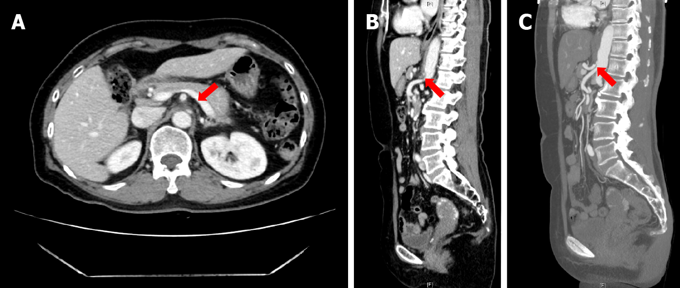
Figure 2 Computed tomography shows the proximal celiac axis narrowing with collateral vessel formation.
A: Axial view; B: Sagittal view; C: Computed tomography angiography confirmed the diagnosis.
- Citation: Kim JE, Rhee PL. Median arcuate ligamentum syndrome: Four case reports. World J Clin Cases 2022; 10(6): 1991-1997
- URL: https://www.wjgnet.com/2307-8960/full/v10/i6/1991.htm
- DOI: https://dx.doi.org/10.12998/wjcc.v10.i6.1991


