Copyright
©The Author(s) 2022.
World J Clin Cases. Feb 26, 2022; 10(6): 1946-1951
Published online Feb 26, 2022. doi: 10.12998/wjcc.v10.i6.1946
Published online Feb 26, 2022. doi: 10.12998/wjcc.v10.i6.1946
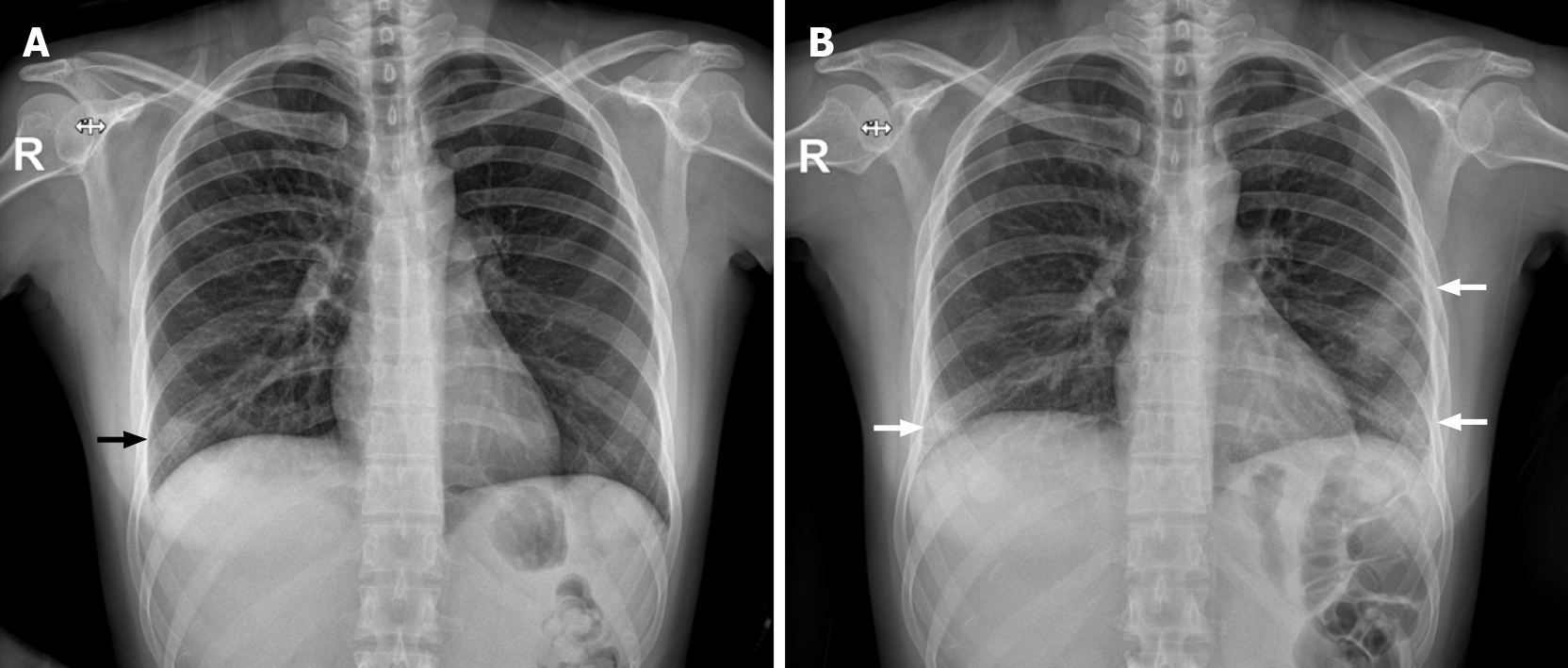
Figure 1 Chest radiography findings.
A: Ill-defined increased opacities in the right lower lobe at first onset (black arrow); B: Increased bilateral patchy opacities in both lower lobes with a subpleural portion at second onset (white arrow).
- Citation: Lee YJ, Kim YS. Cryptogenic organizing pneumonia associated with pregnancy: A case report. World J Clin Cases 2022; 10(6): 1946-1951
- URL: https://www.wjgnet.com/2307-8960/full/v10/i6/1946.htm
- DOI: https://dx.doi.org/10.12998/wjcc.v10.i6.1946


