Published online Feb 6, 2022. doi: 10.12998/wjcc.v10.i4.1326
Peer-review started: August 1, 2021
First decision: November 7, 2021
Revised: November 12, 2021
Accepted: December 22, 2021
Article in press: December 22, 2021
Published online: February 6, 2022
Processing time: 176 Days and 0.4 Hours
In the clinical treatment of diseases related to ureteral duplication, it is very important to make a clear diagnosis before surgery because different types of ureteral duplication correspond to different treatment options. Inverted Y ureteral duplication with ectopic ureters and multiple urinary calculi is clinically rare. This case can help clinicians increase their understanding of this disease and gain some experience in its diagnosis and treatment.
A 36-year-old male who was previously healthy presented to the hospital with lumbar pain. Percussion of the right kidney area showed the patient had pain. Computed tomography scans revealed multiple urinary calculi in the right urinary system. Computed tomography urography revealed a duplicated ureteral malformation with an ectopic ureter. A transurethral ureteroscopic holmium laser lithotripsy was performed successfully. Intraoperative retrograde ureterography was performed, and the ectopic ureter was visible. We informed the family of the intraoperative findings and suggested laparoscopic ectopic ureterectomy for the ectopic ureteral stones. Unfortunately, the family temporarily refused laparoscopic surgery. The patient did not feel any discomfort after one year of follow-up.
Inverted Y ureteral duplication with an ectopic ureter and multiple urinary calculi is rare. Clinicians must be highly vigilant, make a correct diagnosis before surgery, determine the type of ureteral duplication and the distribution of urinary calculi, and then draw up a reasonable treatment plan to avoid unnecessary complications.
Core Tip: The inverted Y duplication ureteral malformation with ectopic ureters and multiple urinary calculi is clinically rare. The diagnosis should be made correctly before operation. It is important to determine the type of ureteral malformation and the distribution of urinary calculi. Endoscopic lithotripsy combined with laparoscopic surgery is a good method for treatment.
- Citation: Ye WX, Ren LG, Chen L. Inverted Y ureteral duplication with an ectopic ureter and multiple urinary calculi: A case report. World J Clin Cases 2022; 10(4): 1326-1332
- URL: https://www.wjgnet.com/2307-8960/full/v10/i4/1326.htm
- DOI: https://dx.doi.org/10.12998/wjcc.v10.i4.1326
Ureteral duplication is defined as two ureters in the ipsilateral urinary system. A ureter with an abnormal positioning is called an ectopic ureter, and it often has an orifice outside the bladder triangle in duplicated ureteral malformations. Inverted Y duplicated ureteral malformation with ectopic ureteral calculi is uncommon in the literature, and only a few cases have been reported[1]. There are no relevant reports on cases with multiple urinary calculi in the renal pelvis and ureters.
A 36-year-old man was admitted to the hospital with complaints of right lumbar pain for one month.
The patient saw a doctor due to right lumbar pain for one month. He had no gross hematuria, chills, fever, or urinary incontinence. No history of trauma was reported.
The patient had no notable previous medical history.
The patient denied any family history and had no notable history.
The patient was conscious. His vital signs were as follows: body temperature, 36.5 °C; pulse, 82/min; respiration rate, 18/min; blood pressure, 130/72 mmHg; right kidney area percussion pain was positive; and abdominal findings were unremarkable.
Laboratory data were unremarkable except for urine red blood cells (+++) on urinalysis.
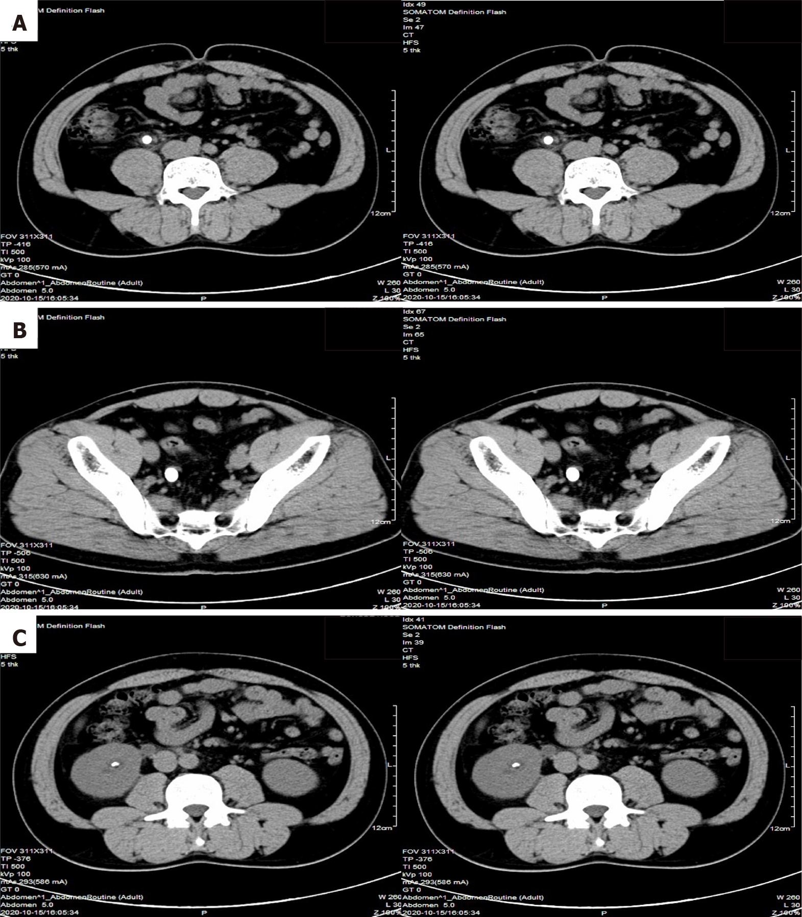
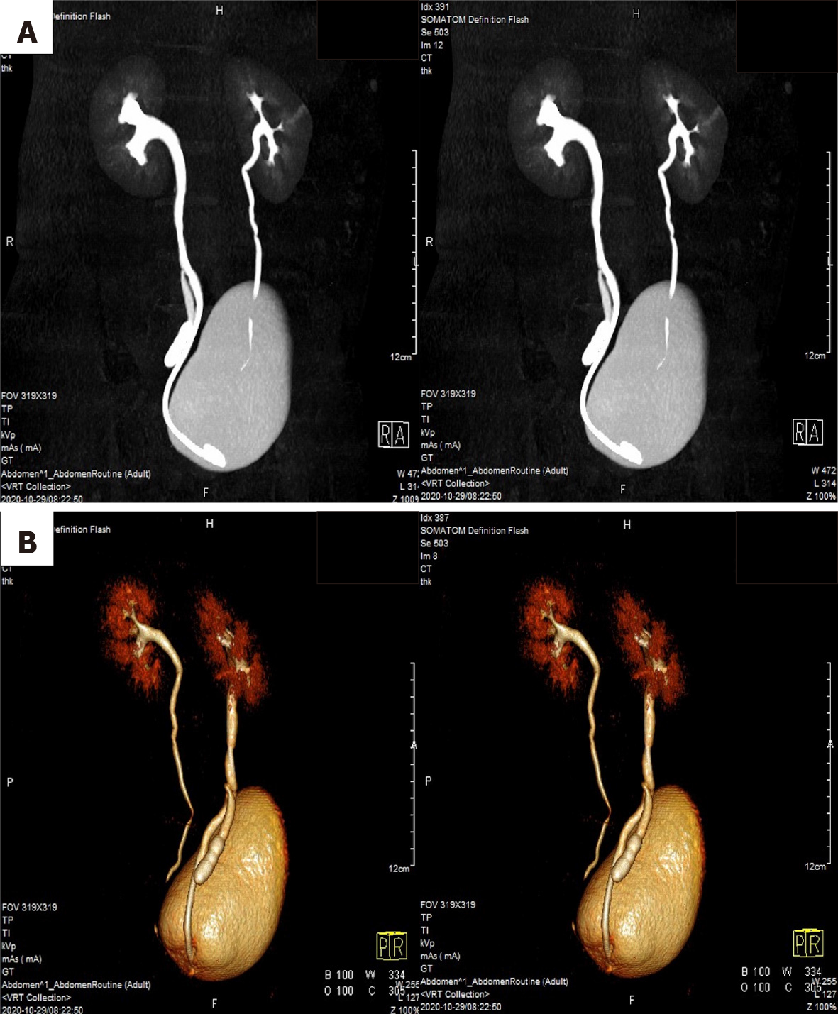
Computed tomography scans revealed one stone in the right upper ureter, multiple stones in the right lower ureter, and other stones in the right kidney (Figure 1). Computed tomography urography and three-dimensional reconstructions revealed that the middle of the right ureter was divided into two branches, one ureter descended to the bladder with a normal bladder opening and the other ureter descended to the vicinity of the right seminal vesicle with an ectopic opening, suggesting duplicated ureteral malformation with an ectopic ureter (Figure 2).
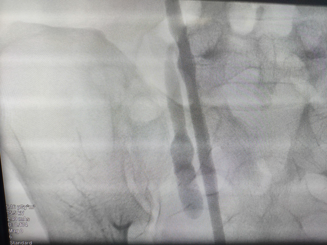
From the imaging findings and intraoperative retrograde ureterography, the final diagnosis was as follows: A right inverted Y duplicated ureteral malformation; right fusion broncho-ureteral stones; multiple stones in the ectopic ureter; and a right kidney stone (Figure 3).
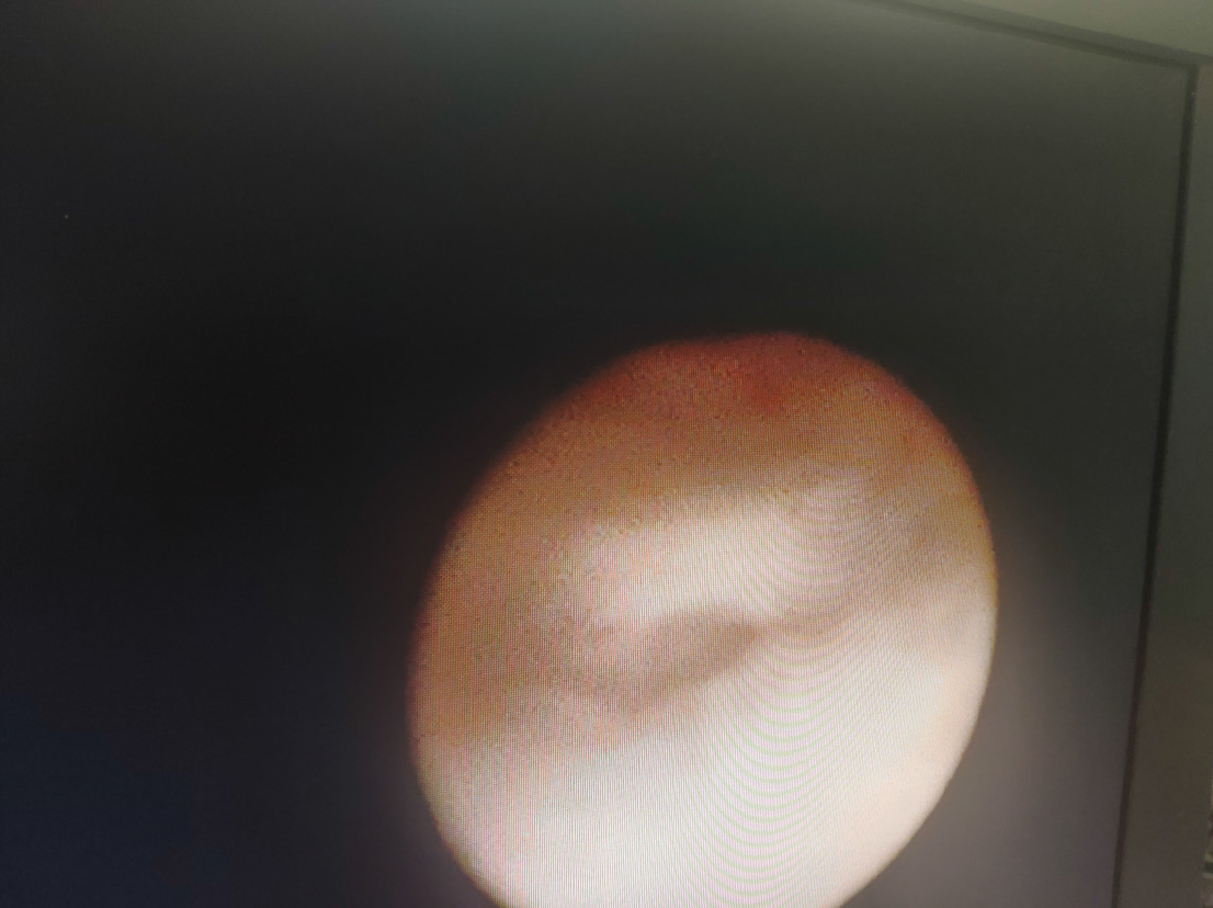
Excluding the operative contraindications, a right transurethral ureteroscopic holmium laser lithotripsy was performed on November 10, 2020. No ectopic ureteral opening was found in the urethra or bladder by endoscopy, and ureteroscopy was used to enter the right ureter through a normal bladder opening along the guidewire. There was a stone in the upper ureter, approximately 1.5 cm × 1.0 cm in size, and a stone was present in the lower calyx of the right kidney, approximately 1.0 cm × 1.0 cm in size. A holmium laser was used to pulverize the stones. The ureteral sheath was placed into the right ureter, and the contrast agent was injected. The ectopic ureter was observed by fluoroscopy using a C model arm X-ray machine. A duplicated ureteral bifurcation could be observed with a flexible ureteroscope (Figure 4), and the intraoperative diagnosis was an inverted Y ureteral duplication and ectopic ureteral multiple stones. We informed the patient’s family members regarding the intraoperative findings and recommended that he undergo laparoscopic ectopic ureterectomy. Unfortunately, the family temporarily refused laparoscopic surgery.
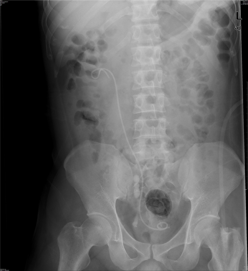
The KUB was rechecked on the second day after surgery (Figure 5), and then the patient was discharged. The double J tube was removed 3 wk after the operation. The patient did not feel any discomfort after one year of follow-up.
Inverted Y ureteral duplication is a rare congenital malformation of the ureter. There are a few reports in the literature. This malformation occurs when two distal ureters fuse into one and then enter the renal pelvis. Similar to other types of duplicated ureteral malformations, the incidence of inverted Y ureteral duplication in women is higher than that in men[2]. In inverted Y ureteral duplication, two distal ureters can open into the bladder. Usually, one ureter opens into the bladder, and the other ureter has an ectopic opening or the end of the ureter has atresia. The mechanism of its formation is not clear, but it is currently thought that its formation is related to the fusion of two independent ureteral bud tips into one tube before connecting to the metanephros[3].
The clinical symptoms of patients with an inverted Y duplicated ureteral malformation are diverse, and they mainly depend on the location of the ectopic ureteral opening. The opening of ectopic ureters into the vulva or reproductive organs can cause incontinence[4,5]; the opening of ectopic ureters into the bladder and form ureteral cysts can cause bladder outlet obstruction[6]; the opening of ectopic ureters into the seminal vesicles can cause discomfort in the lower abdomen or infertility[7]; and ureteral stones in the ectopic ureter may cause abdominal pain or gross hematuria[1].
An inverted Y duplicated ureteral malformation cannot be diagnosed by clinical symptoms and is mainly diagnosed by ureterography. Retrograde ureterography routinely develops by X-rays and can also develop by contrast-enhanced ultrasound[8].
There is no standard for the treatment of inverted Y duplicated ureteral malformations or their comorbidities. Asymptomatic patients with inverted Y duplicated ureteral malformations can be followed up for observation. For comorbidities caused by ectopic ureters, corresponding treatment can be performed. When ectopic ureteral cysts cause bladder outflow obstruction, removal of the endoscopic cyst is feasible[9]. For ectopic ureters that cause incontinence or intraureteral stones, laparoscopic ectopic ureterectomy is feasible[10], and replantation of the ureter can also be considered[1].
In this case, the inverted Y ureteral duplication combined with multiple urinary calculi not only contributed to the stones in the renal pelvis and the fused broncho-ureter, but also contributed to the development of multiple stones in the ectopic ureter. It is not clear whether the ectopic ureteral stones developed from the kidney or another primary site. Fused broncho-ureteral stones can cause hydronephrosis, which can manifest as lumbar pain, while ectopic ureteral stones may not cause clinical symptoms and are often ignored. The methods for treating urinary calculi in different parts of the urinary tract are different. In this case, a transurethral ureteroscopic holmium laser lithotripsy was used to treat fused broncho-ureteral calculi and intrarenal pelvic calculi, and the results were satisfactory. Our only regret is that the patient refused to treat the ectopic ureteral stones, and surgical specimens could not be obtained. During the one-year follow-up, the patient had no obvious symptoms, such as lumbar pain. However, because of long-term entrapment and stimulation by urinary calculi, urinary calculi can cause urinary tract infections and can increase the risk of cancer, and it is recommended that patients with an ectopic ureter have it removed as soon as possible during follow-up. The most minimally invasive and effective method currently reported in the literature is laparoscopic ectopic ureterectomy.
For patients with an inverted Y ureteral duplication and multiple urinary calculi, it is important to clarify the type of ureteral malformation and the location of the stones, and these identifications can have a positive effect on the treatment. For stones in the renal pelvis and ureters with normal openings, endoscopic lithotripsy can be used to achieve good results. For stones in an ectopic ureter, it is difficult to find or locate the ectopic ureteral opening, and most patients are advised to have the ectopic ureter removed. At present, laparoscopic ectopic ureterectomy is a common procedure, and the consequent outcome is usually good.
Provenance and peer review: Unsolicited article; Externally peer reviewed.
Peer-review model: Single blind
Specialty type: Urology and nephrology
Country/Territory of origin: China
Peer-review report’s scientific quality classification
Grade A (Excellent): 0
Grade B (Very good): B
Grade C (Good): C, C, C
Grade D (Fair): 0
Grade E (Poor): E
P-Reviewer: Beraldo RF, Corvino A, Islam SMRU, Iwao Y S-Editor: Liu JH L-Editor: A P-Editor: Liu JH
| 1. | Masuda H, Yamada T, Nagamatsu H, Nagahama K, Negishi T. [Inverted Y ureteral duplication with a ureteral stone in atretic segment: a case report]. Hinyokika Kiyo. 1994;40:423-425. [PubMed] |
| 2. | Klauber GT, Reid EC. Inverted Y reduplication of the ureter. J Urol. 1972;107:362-364. [RCA] [PubMed] [DOI] [Full Text] [Cited by in Crossref: 18] [Cited by in RCA: 18] [Article Influence: 0.3] [Reference Citation Analysis (0)] |
| 3. | Roy Choudhury S, Chadha R, Bagga D, Puri A, Debnath PR. Spectrum of ectopic ureters in children. Pediatr Surg Int. 2008;24:819-823. [RCA] [PubMed] [DOI] [Full Text] [Cited by in Crossref: 33] [Cited by in RCA: 27] [Article Influence: 1.6] [Reference Citation Analysis (0)] |
| 4. | Shimura H, Mitsui T, Aoki T, Kamiyama M, Yamagishi T, Takeda M. Persistent Urinary Incontinence After Nephrectomy: A Case of Inverted-Y Ureteral Duplication with Ectopic Ureteral Insertion into the Vagina. Urol Case Rep. 2016;9:58-61. [RCA] [PubMed] [DOI] [Full Text] [Full Text (PDF)] [Cited by in Crossref: 2] [Cited by in RCA: 2] [Article Influence: 0.2] [Reference Citation Analysis (0)] |
| 5. | Souffrant D, Hagerty J, Colon-Sanchez K, Epelman M, Ellsworth P. Intermittent Urinary Incontinence Secondary to Inverted-Y Ureteral Duplication With Perianal Ectopia. Urology. 2019;127:124-126. [RCA] [PubMed] [DOI] [Full Text] [Cited by in Crossref: 2] [Cited by in RCA: 1] [Article Influence: 0.2] [Reference Citation Analysis (0)] |
| 6. | Suzuki H, Yanagi S, Namiki T, Takano M. [A case of inverted Y ureteral duplication with an ectopic ureterocele]. Nihon Hinyokika Gakkai Zasshi. 1992;83:1713-1716. [RCA] [PubMed] [DOI] [Full Text] [Cited by in Crossref: 1] [Cited by in RCA: 1] [Article Influence: 0.0] [Reference Citation Analysis (0)] |
| 7. | Schenkman NS, Fernandez EB, Wind G, Irby P. Inverted Y duplication of the ureter associated with a distal limb ectopic to the seminal vesicle. J Urol. 1994;152:946-947. [RCA] [PubMed] [DOI] [Full Text] [Cited by in Crossref: 7] [Cited by in RCA: 8] [Article Influence: 0.3] [Reference Citation Analysis (0)] |
| 8. | Usawachintachit M, Tzou DT, Mongan J, Taguchi K, Weinstein S, Chi T. Feasibility of Retrograde Ureteral Contrast Injection to Guide Ultrasonographic Percutaneous Renal Access in the Nondilated Collecting System. J Endourol. 2017;31:129-134. [RCA] [PubMed] [DOI] [Full Text] [Cited by in Crossref: 6] [Cited by in RCA: 6] [Article Influence: 0.7] [Reference Citation Analysis (0)] |
| 9. | Regan TC, Dejter SW Jr. Inverted Y duplication of the ureter with associated ureterocele and bladder outlet obstruction. J Urol. 1996;155:642. [PubMed] |
| 10. | Upadhyay J, Zuckerman JM, Shekarriz B. Minimally invasive surgical approach to excision of symptomatic ectopic inverted-Y ureteral duplications. J Pediatr Urol. 2013;9:e114-e116. [RCA] [PubMed] [DOI] [Full Text] [Cited by in Crossref: 6] [Cited by in RCA: 6] [Article Influence: 0.5] [Reference Citation Analysis (0)] |


