Copyright
©The Author(s) 2022.
World J Clin Cases. Dec 26, 2022; 10(36): 13157-13166
Published online Dec 26, 2022. doi: 10.12998/wjcc.v10.i36.13157
Published online Dec 26, 2022. doi: 10.12998/wjcc.v10.i36.13157
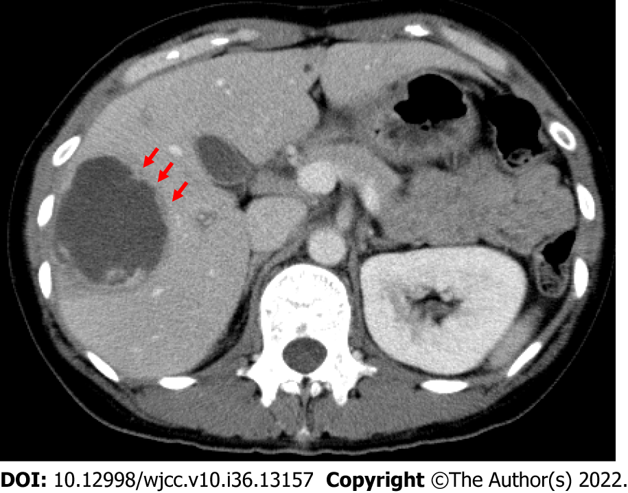
Figure 1 Computed tomography of a 44-year-old woman with a type I abscess.
The axial computed tomography image illustrates the non-enhancing and ragged edge of the abscess in the absence of a definite wall, peripheral septa, and ragged edges; these edges exhibited both irregular and interrupted enhancement (arrows).
- Citation: Usuda D, Tsuge S, Sakurai R, Kawai K, Matsubara S, Tanaka R, Suzuki M, Takano H, Shimozawa S, Hotchi Y, Tokunaga S, Osugi I, Katou R, Ito S, Mishima K, Kondo A, Mizuno K, Takami H, Komatsu T, Oba J, Nomura T, Sugita M. Amebic liver abscess by Entamoeba histolytica. World J Clin Cases 2022; 10(36): 13157-13166
- URL: https://www.wjgnet.com/2307-8960/full/v10/i36/13157.htm
- DOI: https://dx.doi.org/10.12998/wjcc.v10.i36.13157


