Published online Dec 26, 2022. doi: 10.12998/wjcc.v10.i36.13451
Peer-review started: September 26, 2022
First decision: October 28, 2022
Revised: November 10, 2022
Accepted: December 5, 2022
Article in press: December 5, 2022
Published online: December 26, 2022
Processing time: 91 Days and 4 Hours
The clinical course of acute myocarditis ranges from the occurrence of a few symptoms to the development of fatal fulminant myocarditis. Specifically, fulminant myocarditis causes clinical deterioration very rapidly and aggressively. The long-term prognosis of myocarditis is varied, and it fully recovers without leaving any special complications. However, even after recovery, heart failure may occur and eventually progress to dilated cardiomyopathy (DCM), which causes serious left ventricular dysfunction. In the case of follow-up observation, no clear guidelines have been established.
We report the case of a 21-year-old woman who presented with dyspnea. She became hemodynamically unstable and showed sustained fatal arrhythmias with decreased heart function. She was clinically diagnosed with fulminant myocarditis based on her echocardiogram and cardiac magnetic resonance results. After 2 d, she was readmitted to the emergency department under cardiopulmonary resuscitation and received mechanical ventilation and extracorporeal membrane oxygenation. An implantable cardioverter defibrillator was inserted for secondary prevention. She recovered and was discharged. Prior to being hospitalized for sudden cardiac function decline and arrhythmia, she had been well for 7 years without any complications. She was finally diagnosed with dilated cardiomyopathy.
DCM may develop unexpectedly in patients who have been cured of acute fulminant myocarditis and have been stable with a long period of remission. Therefore, they should be carefully and regularly observed clinically throughout long-term follow-up.
Core Tip: While dilated cardiomyopathy (DCM) has been well-known as a complication in patients who develop fulminant myocarditis, it is still unclear when DCM might occur. We report the case of a young woman who developed DCM after 7 years of remission without any special complications after recovering from viral myocarditis. No case of DCM development has been reported after such a long latent period of normal cardiac function after a full recovery from viral fulminant myocarditis. We, therefore, suggest that clinicians be aware that DCM can develop unexpectedly and that careful clinical monitoring is required regularly.
- Citation: Lee SD, Lee HJ, Kim HR, Kang MG, Kim K, Park JR. Development of dilated cardiomyopathy with a long latent period followed by viral fulminant myocarditis: A case report. World J Clin Cases 2022; 10(36): 13451-13457
- URL: https://www.wjgnet.com/2307-8960/full/v10/i36/13451.htm
- DOI: https://dx.doi.org/10.12998/wjcc.v10.i36.13451
Myocarditis, an inflammation of the myocardium, is also defined as inflammatory cardiomyopathy characterized by worsening cardiac dysfunction and cardiac remodeling in terms of the size, shape, and structure of the heart[1]. Etiologies of acute myocarditis include various infections, autoimmune diseases, hypersensitivity reactions, and toxic reactions to drugs and toxins[1]. Clinical presentations of myocarditis vary from asymptomatic or with mild symptoms of chest pain, dyspnea, or palpitations to high-risk cardiac conditions with severe heart failure (HF), refractory arrhythmias, cardiogenic shock, and sudden cardiac death[2].
Regarding the severity and prognosis of myocarditis, most patients successfully recover, and the clinical course is mostly self-limited. However, in a study, nearly 30% of individuals reportedly developed dilated cardiomyopathy (DCM)[3]. Furthermore, prompt and aggressive treatment can help achieve complete recovery in patients who develop a fulminant presentation with severe left ventricular (LV) dysfunction[4]. Although studies have found that patients with myocarditis have poor long-term clinical outcomes[5-7], the outcomes and follow-up strategies after full recovery require further investigation.
Herein, we present a case of delayed development of DCM after a long remission period of fulminant myocarditis.
A 21-year-old woman who presented with dyspnea, tachycardia, and hypotension was transferred to our emergency department.
She was admitted to the local hospital for 2 d for upper abdominal discomfort and constipation.
She had a history of cesarean delivery a year before.
She had no history of family history of any heart disease.
Her initial vital signs were as follows: Blood pressure of 137/67 mmHg, pulse rate of 184 beats per min, respiration of 40 breaths per minute, and a body temperature of 36.7 ℃. Other general examinations were not remarkable. Atrial fibrillation was detected, and the rate still increased even after the administration of digitalis. Her systolic blood pressure suddenly dropped, so she received electric defibrillation at 100 J, and her hemodynamic status was stabilized with a sinus heart rhythm.
Remarkable laboratory findings were highly elevated levels of liver enzymes, i.e., aspartate aminotransferase of 3381 U/L (reference, < 37 U/L) and alanine aminotransferase of 1197 U/L (reference, < 41 U/L); extremely increased B-type natriuretic peptide level of 2233 pg/mL (reference range, 0-100 pg/mL); mild hypokalemia, with serum potassium concentration of 3.2 mmol/L (reference range, 3.3-5.1 mmol/L); white blood cell count of 11710 × 103/mm3; and C-reactive protein of 10.7 mg/L. Other than that, no abnormal findings, including cardiac enzymes, were found.
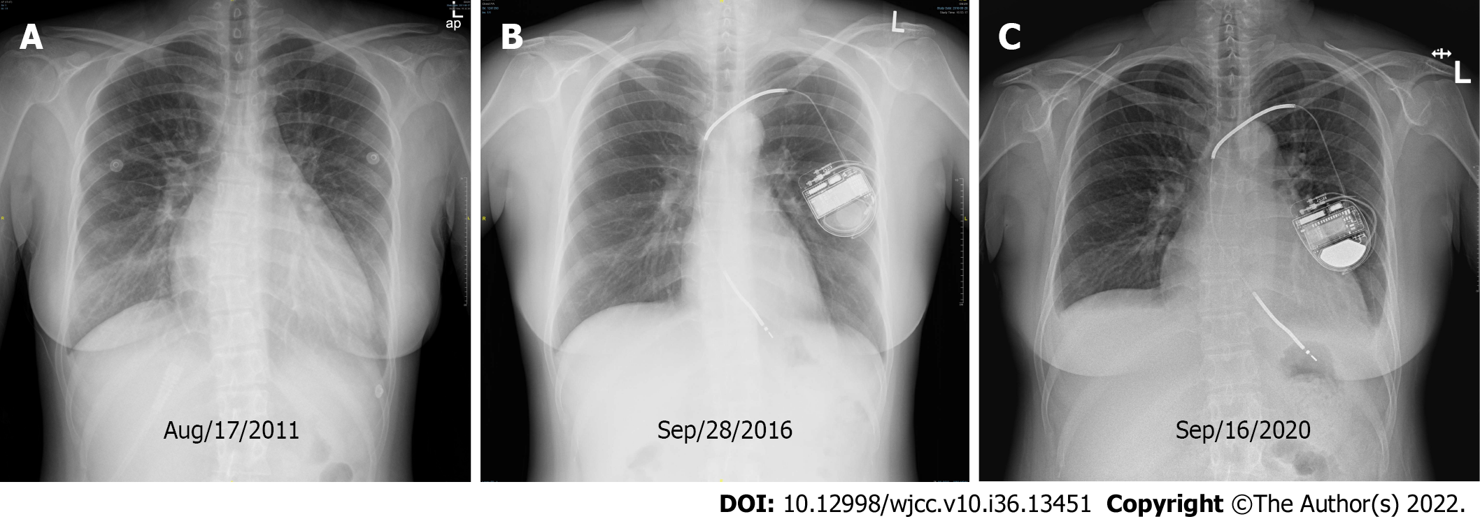
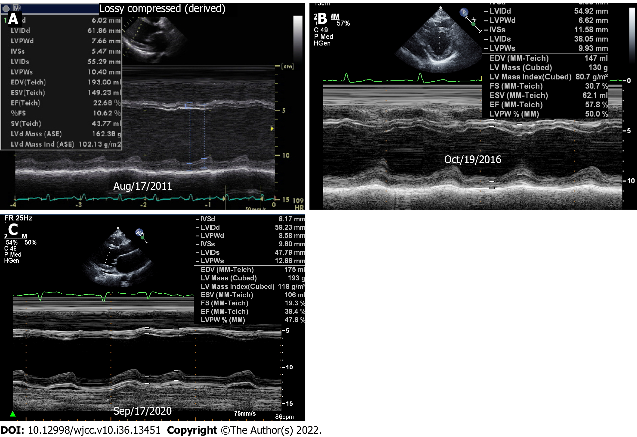
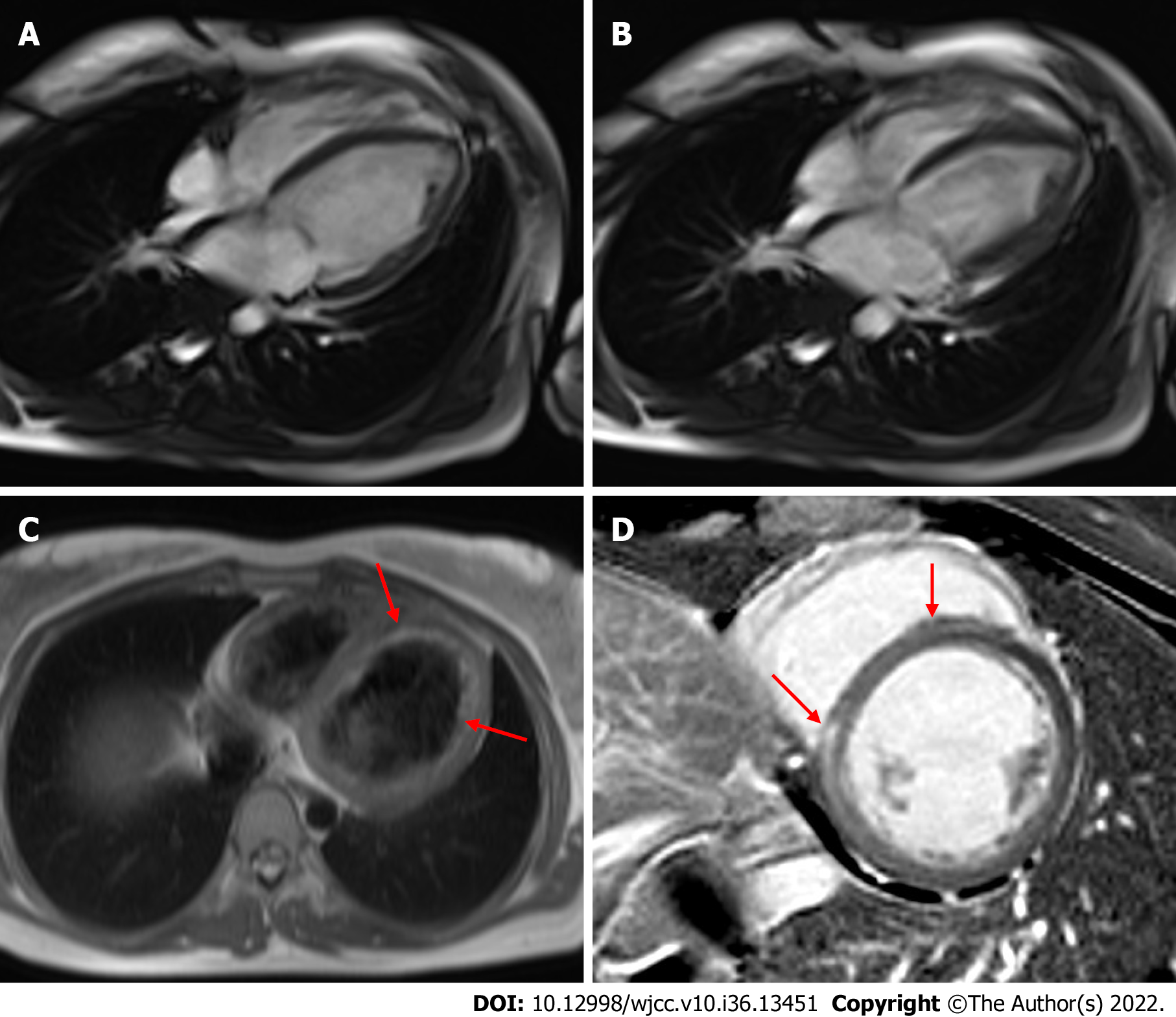
Her initial chest X-ray imaging showed remarkable cardiomegaly and bilateral pulmonary edema (Figure 1A). The initial transthoracic echocardiography (TTE) demonstrated dilated left atrium and LV and severe LV systolic dysfunction with an ejection fraction (EF) of 24% (Figure 2A; Supplementary Video 1). Mild mitral regurgitation and a small amount of pericardial effusion were found. On day 7 of hospitalization, cardiac magnetic resonance (CMR) imaging revealed endocardial edematous change and delayed gadolinium enhancement from the interventricular septum to the inferior wall of the LV (Figure 3). The myocardial perfusion of contrast was normal on CMR. Liver ultrasonography to rule out severe hepatic illness revealed only mild-to-moderate fatty liver.
She was diagnosed with acute myocarditis based on her acute clinical presentation and CMR findings. Increased liver enzymes improved dramatically with diuretic therapy and were thought to be the result of hepatic congestion following HF. After clinical stabilization, she was discharged and outpatient follow-up was arranged. However, she was readmitted at night of the same day that cardiopulmonary resuscitation was performed. On arrival, endotracheal intubation was performed, and repetitive defibrillation continued throughout the day. Severe arrhythmias, including ventricular fibrillation, ventricular tachycardia, and supraventricular tachycardia, recurred with hypotension. Subsequently, amiodarone and dobutamine were infused, and extracorporeal membrane oxygenation (ECMO) was initiated for circulatory support. As a result, her vital sign stabilized, and fatal arrhythmia was not observed. ECMO was discontinued, and an implantable cardioverter defibrillator (ICD) was inserted for secondary prevention.
On the second visit to the emergency department, the levels of cardiac enzymes were increased: Creatinine kinase-MB, 35.4 ng/mL (reference, < 5 ng/mL) and troponin-I, 2.67 ng/mL (reference range, 0-0.04 ng/mL). High neutralizing antibody titers to Coxsackie B1 virus (titer 1:64) were confirmed, and autoantibodies were negative. Although endomyocardial biopsy was not performed, she was clinically diagnosed with fulminant myocarditis based on TTE and CMR findings on clinical presentation. For HF, beta blockers, angiotensin receptor blockers, and diuretics were prescribed. TTE on day 21 revealed improved EF up to 40%.
One year later, the cardiomegaly detected by chest X-ray imaging was improved, and her follow-up LVEF recovered to normal range (Figures 1B and 2B; Supplementary Video 2). In the 7-year follow-up outpatient care, her condition and ICD without medications were regularly checked up. During the follow-up, she did not present any symptoms or signs, and there was only one event of inappropriate shock as a response to sinus tachycardia.
On the 8th year, paroxysmal atrial fibrillation and rapid ventricular response were detected by ICD recording, and the LVEF decreased to 42%, which means that she had a relapse of HF (Figures 1C and 2C; Supplementary Video 3). There was no definite evidence of aggravating factors regarding her disease such as medication, alcohol, infection, and even emotional stress. She presented dry cough, ankle edema, and chest discomfort gradually. Guideline-based medications were started again, and her condition symptomatically improved.
Initially, the patient was clinically diagnosed with fulminant myocarditis based on TTE and CMR findings on clinical presentation. The patient was finally diagnosed with DCM with a long remission period after viral fulminant myocarditis.
She was given an angiotensin receptor blocker, a vitamin K antagonist, and digitalis as conventional treatment for DCM and atrial fibrillation.
The most recent imaging performed on March 2021 showed a slight improvement in LVEF (47%). She is currently not experiencing any clinical adverse events.
Fulminant myocarditis is a severe inflammatory myocardial disease often induced by cardiotropic viruses such as parvovirus, human herpes virus-6, coxsackie virus, human immunodeficiency virus, and cytomegalovirus[5]. Despite different clinical courses and outcomes, up to 30% of patients with acute myocarditis progressed to DCM, which is clinically defined by LV dilatation and systolic dysfunction[3]. The long-term poor clinical outcomes of patients with myocarditis have been reported[5,6], Escher et al[7] reported that about 50% of patients who suffered from acute myocarditis developed diastolic HF with preserved EF in a 6-year long-term follow-up period. However, it is unknown when DCM develops following the resolution of fulminant myocarditis.
In this case, we believed the patient had recovered completely from fulminant myocarditis, and she had been well with normal LVEF and normal diastolic function for 7 years. The paroxysmal atrial fibrillation event was the first sign of recurrent HF. It took 6 mo from the event of atrial fibrillation on the ICD to the onset of symptomatic HF. Studies and cases of recurrent myocarditis have been reported, with different intervals and varying causes[8,9]. However, we believe that our case is consistent with the study of Escher et al[7] in that the patient developed a long-latency relapse of HF because her symptoms and signs aggravated gradually. Recently, inflammatory cardiomyopathy, including DCM following acute myocarditis, has been recognized. Latent myocarditis caused by persistent virus and chronic inflammation is the major pathology of inflammatory cardiomyopathy, and it is commonly reported as a chronic and progressive impairment[10,11]. In another aspect, Spotnitz and Lesch[12] hypothesized the idiopathic dilated cardiomyopathy could be a late complication of healed viral myocarditis. Ghanizada et al[5] demonstrated a higher risk of HF hospitalization and all-cause mortality among patients with myocarditis, even in patients without cardiovascular events and those on HF medications within the first year of discharge, compared to matched controls.
As DCM has been demonstrated to be one of the most common hereditary cardiomyopathies, genetic analysis of mutations for DCM became essential to determine the pathogenesis of the disease[13]. Our patient did not participate in the genetic test for cardiomyopathies because she had no family history related to cardiovascular disease and her medical history of myocarditis was apparent. However, further DCM genetic evaluation would be helpful to rule out any genetic cause for the development of DCM.
To the best of our knowledge, there has been no reported case of DCM developing after such a long latent period while maintaining normal LV function after a full recovery from viral fulminant myocarditis. To date, the long-term follow-up strategy after restoration of fulminant myocarditis has not been established. Moreover, the TRED-HF trial demonstrated a relapse of HF among patients who stopped pharmacological treatment after their LV function was restored[14]. Therefore, we suggest that continuous and regular follow-up and individualized pharmacologic treatment would be required in patients recovering from acute myocarditis. This precaution also should be taken in the case of COVID-19 myocarditis, which has been issued for a severe cardiovascular complication, as well as in some cases of COVID-19 mRNA vaccine-associated myocarditis.
We presented one clinical experience that even though patients who are thought to be fully recovered from acute fulminant myocarditis and seemed to be stable for a long-time follow-up period, may develop DCM unexpectedly and they should be clinically monitored at outpatient clinic carefully and regularly with a long period of follow-up.
Provenance and peer review: Unsolicited article; Externally peer reviewed.
Peer-review model: Single blind
Specialty type: Cardiac and cardiovascular systems
Country/Territory of origin: South Korea
Peer-review report’s scientific quality classification
Grade A (Excellent): 0
Grade B (Very good): 0
Grade C (Good): C
Grade D (Fair): D
Grade E (Poor): 0
P-Reviewer: Dai HL, China; Feng R, China S-Editor: Chen YL L-Editor: A P-Editor: Chen YL
| 1. | Tschöpe C, Ammirati E, Bozkurt B, Caforio ALP, Cooper LT, Felix SB, Hare JM, Heidecker B, Heymans S, Hübner N, Kelle S, Klingel K, Maatz H, Parwani AS, Spillmann F, Starling RC, Tsutsui H, Seferovic P, Van Linthout S. Myocarditis and inflammatory cardiomyopathy: current evidence and future directions. Nat Rev Cardiol. 2021;18:169-193. [RCA] [PubMed] [DOI] [Full Text] [Full Text (PDF)] [Cited by in Crossref: 271] [Cited by in RCA: 703] [Article Influence: 140.6] [Reference Citation Analysis (0)] |
| 2. | Ammirati E, Veronese G, Bottiroli M, Wang DW, Cipriani M, Garascia A, Pedrotti P, Adler ED, Frigerio M. Update on acute myocarditis. Trends Cardiovasc Med. 2021;31:370-379. [RCA] [PubMed] [DOI] [Full Text] [Full Text (PDF)] [Cited by in Crossref: 60] [Cited by in RCA: 81] [Article Influence: 20.3] [Reference Citation Analysis (0)] |
| 3. | Lampejo T, Durkin SM, Bhatt N, Guttmann O. Acute myocarditis: aetiology, diagnosis and management. Clin Med (Lond). 2021;21:e505-e510. [RCA] [PubMed] [DOI] [Full Text] [Cited by in Crossref: 55] [Cited by in RCA: 87] [Article Influence: 21.8] [Reference Citation Analysis (0)] |
| 4. | Gupta S, Markham DW, Drazner MH, Mammen PP. Fulminant myocarditis. Nat Clin Pract Cardiovasc Med. 2008;5:693-706. [RCA] [PubMed] [DOI] [Full Text] [Cited by in Crossref: 136] [Cited by in RCA: 143] [Article Influence: 8.4] [Reference Citation Analysis (0)] |
| 5. | Ghanizada M, Kristensen SL, Bundgaard H, Rossing K, Sigvardt F, Madelaire C, Gislason GH, Schou M, Hansen ML, Gustafsson F. Long-term prognosis following hospitalization for acute myocarditis - a matched nationwide cohort study. Scand Cardiovasc J. 2021;55:264-269. [RCA] [PubMed] [DOI] [Full Text] [Cited by in Crossref: 1] [Cited by in RCA: 9] [Article Influence: 2.3] [Reference Citation Analysis (0)] |
| 6. | Chang JJ, Lin MS, Chen TH, Chen DY, Chen SW, Hsu JT, Wang PC, Lin YS. Heart Failure and Mortality of Adult Survivors from Acute Myocarditis Requiring Intensive Care Treatment - A Nationwide Cohort Study. Int J Med Sci. 2017;14:1241-1250. [RCA] [PubMed] [DOI] [Full Text] [Full Text (PDF)] [Cited by in Crossref: 37] [Cited by in RCA: 44] [Article Influence: 5.5] [Reference Citation Analysis (0)] |
| 7. | Escher F, Westermann D, Gaub R, Pronk J, Bock T, Al-Saadi N, Kühl U, Schultheiss HP, Tschöpe C. Development of diastolic heart failure in a 6-year follow-up study in patients after acute myocarditis. Heart. 2011;97:709-714. [RCA] [PubMed] [DOI] [Full Text] [Cited by in Crossref: 56] [Cited by in RCA: 48] [Article Influence: 3.4] [Reference Citation Analysis (0)] |
| 8. | Karavidas A, Lazaros G, Noutsias M, Matzaraki V, Danias PG, Pyrgakis V, Voudris V, Adamopoulos S. Recurrent coxsackie B viral myocarditis leading to progressive impairment of left ventricular function over 8 years. Int J Cardiol. 2011;151:e65-e67. [RCA] [PubMed] [DOI] [Full Text] [Cited by in Crossref: 9] [Cited by in RCA: 8] [Article Influence: 0.6] [Reference Citation Analysis (0)] |
| 9. | Kytö V, Sipilä J, Rautava P. Rate and patient features associated with recurrence of acute myocarditis. Eur J Intern Med. 2014;25:946-950. [RCA] [PubMed] [DOI] [Full Text] [Cited by in Crossref: 15] [Cited by in RCA: 11] [Article Influence: 1.0] [Reference Citation Analysis (0)] |
| 10. | Lasrado N, Reddy J. An overview of the immune mechanisms of viral myocarditis. Rev Med Virol. 2020;30:1-14. [RCA] [PubMed] [DOI] [Full Text] [Cited by in Crossref: 23] [Cited by in RCA: 84] [Article Influence: 16.8] [Reference Citation Analysis (0)] |
| 11. | Imanaka-Yoshida K. Inflammation in myocardial disease: From myocarditis to dilated cardiomyopathy. Pathol Int. 2020;70:1-11. [RCA] [PubMed] [DOI] [Full Text] [Cited by in Crossref: 38] [Cited by in RCA: 55] [Article Influence: 9.2] [Reference Citation Analysis (0)] |
| 12. | Spotnitz MD, Lesch M. Idiopathic dilated cardiomyopathy as a late complication of healed viral (Coxsackie B virus) myocarditis: historical analysis, review of the literature, and a postulated unifying hypothesis. Prog Cardiovasc Dis. 2006;49:42-57. [RCA] [PubMed] [DOI] [Full Text] [Cited by in Crossref: 29] [Cited by in RCA: 30] [Article Influence: 1.6] [Reference Citation Analysis (0)] |
| 13. | Malakootian M, Bagheri Moghaddam M, Kalayinia S, Farrashi M, Maleki M, Sadeghipour P, Amin A. Dilated cardiomyopathy caused by a pathogenic nucleotide variant in RBM20 in an Iranian family. BMC Med Genomics. 2022;15:106. [RCA] [PubMed] [DOI] [Full Text] [Full Text (PDF)] [Cited by in Crossref: 1] [Cited by in RCA: 7] [Article Influence: 2.3] [Reference Citation Analysis (0)] |
| 14. | Halliday BP, Wassall R, Lota AS, Khalique Z, Gregson J, Newsome S, Jackson R, Rahneva T, Wage R, Smith G, Venneri L, Tayal U, Auger D, Midwinter W, Whiffin N, Rajani R, Dungu JN, Pantazis A, Cook SA, Ware JS, Baksi AJ, Pennell DJ, Rosen SD, Cowie MR, Cleland JGF, Prasad SK. Withdrawal of pharmacological treatment for heart failure in patients with recovered dilated cardiomyopathy (TRED-HF): an open-label, pilot, randomised trial. Lancet. 2019;393:61-73. [RCA] [PubMed] [DOI] [Full Text] [Full Text (PDF)] [Cited by in Crossref: 350] [Cited by in RCA: 415] [Article Influence: 69.2] [Reference Citation Analysis (0)] |


