Published online Dec 6, 2022. doi: 10.12998/wjcc.v10.i34.12799
Peer-review started: October 14, 2022
First decision: October 24, 2022
Revised: October 30, 2022
Accepted: November 8, 2022
Article in press: November 8, 2022
Published online: December 6, 2022
Processing time: 49 Days and 0.3 Hours
As a rare anomaly, congenital absence of the right coronary artery (RCA) occurs during the development of coronary artery. Patients with congenital absence of the RCA often show no clinical symptoms, and this disease is considered benign. The left coronary artery gives blood supply to the whole myocardium. The prevalence of congenital absence of the RCA is approximately 0.024%-0.066%. There are few cases reported as for this disease. In this work, a patient, with congenital absence of the RCA diagnosed by coronary angiography (CAG), was described.
A 41-year-old man arrived at our hospital for treatment, due to the repeated palpitations for a duration of one year. Considering the possibility of coronary heart disease, the patient underwent CAG that indicated the congenital absence of the RCA. Unfortunately, the patient refused to accept computed tomography coronary angiography (CTCA), to further confirm the congenital absence of the RCA.
Single coronary artery is a rare type of coronary artery abnormality, which usually has no obvious clinical manifestations and is considered as a benign disease. CAG is the main means by which congenital absence of the RCA can be diagnosed, and the disease can also be further confirmed by CTCA.
Core Tip: A rare case of congenital absence of the right coronary artery was identified during coronary angiography of a patient.
- Citation: Zhu XY, Tang XH. Congenital absence of the right coronary artery: A case report. World J Clin Cases 2022; 10(34): 12799-12803
- URL: https://www.wjgnet.com/2307-8960/full/v10/i34/12799.htm
- DOI: https://dx.doi.org/10.12998/wjcc.v10.i34.12799
Congenital absence of the right coronary artery (RCA) is a special case of abnormal coronary anatomy. RCA gives blood supply to the right myocardium from the circumflex branch (LCX). These patients are usually found by coronary angiography (CAG) or computed tomography coronary angiography (CTCA), and the case of congenital absence of the RCA reported in this work was diagnosed by CAG.
On April 22, 2022, a 41-year-old man arrived at Jiujiang University Affiliated Hospital for treatment, due to the repeated palpitations with a duration of one year.
The patient had no obvious symptoms of palpitations one year ago, and his current symptoms were accompanied by chest tightness while without chest pain. According to the description of the patient, each attack had no obvious relation with his physical activity, the duration of each attack could be relieved after a few minutes, and no active diagnosis or treatment was accepted.
The patient denied the history of hypertension and diabetes.
The patient denied smoking, drinking history, and family disease history.
The body temperature was 36.6 ºC, the breathing was 18 breaths/min, the blood pressure was 110/72 mmHg, the heart rate was 70 beats/min, and the physical examination was normal.
No obvious abnormality was found in routine blood analysis, biochemistry, hyperthyroidism, cardiac color Doppler ultrasound, thyroid color Doppler ultrasound, routine electrocardiogram, or dynamic electrocardiogram.
The patient had a congenital absence of the RCA.
As for the treatment of RCA, there is still no standardized guideline, so no surgical intervention was given to the patient based on the experience of others and by combining with the results of CAG. The patient was instructed to take aspirin antiplatelet regularly, take atorvastatin for plaque stabilization, and take metoprolol for ventricular rate control. Moreover, the patient was instructed to engage in appropriate physical activity and to keep a healthy lifestyle, for the primary prevention of coronary heart disease.
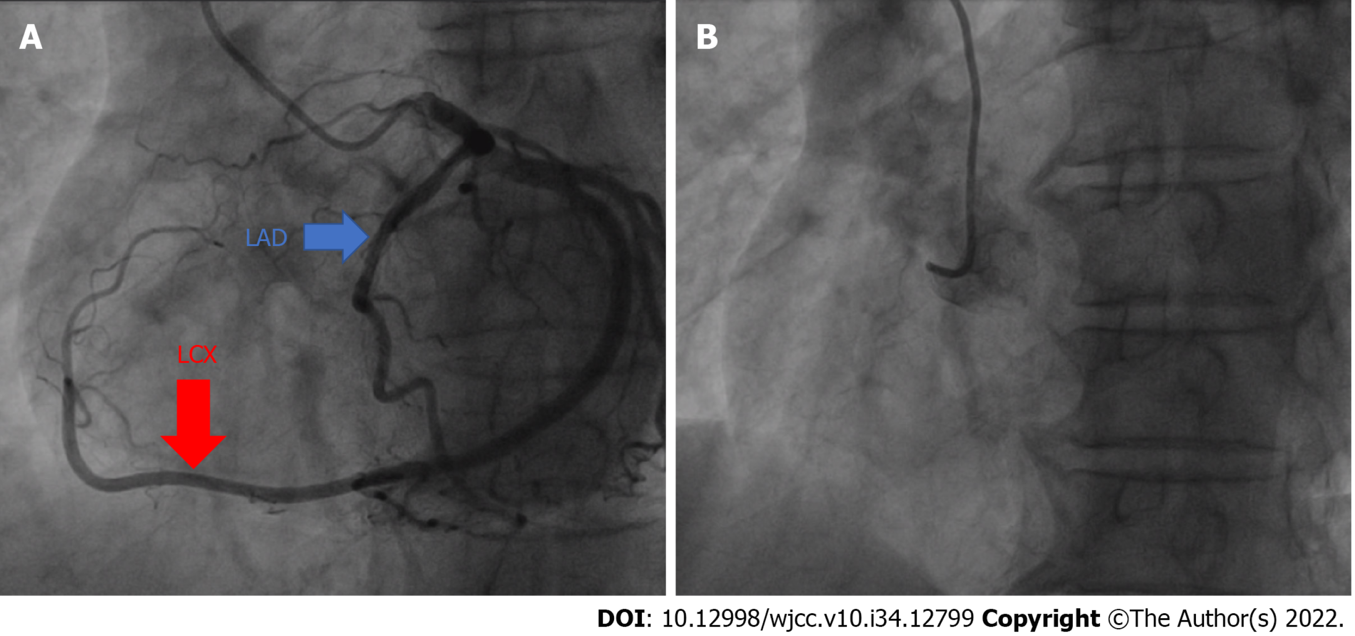
The patient's palpitation symptoms were improved by taking the drug (Metoprolol sustained-release tablets with 47.5 mg/d), and he was discharged from the hospital with a prescription of the drug (Figure 1).
Before discussing coronary artery anomalies, it is necessary to understand the normal anatomy of the coronary arteries[1]. The aortic root consists of three coronary sinuses, namely the left coronary sinus, the right coronary sinus, and the non-coronary sinus. Among them, the left coronary sinus gives rise to the left coronary artery that is divided into the left anterior descending branch and the LCX. While, the right coronary sinus gives rise to the right coronary[2].
Congenital absence of the RCA is a rare abnormal coronary artery disease, with very low incidence in the population, at approximately 0.024%-0.066%[3-7]. Congenital absence of coronary arteries is mostly caused by the defects in coronary artery development during embryonic development[3-6]. Patients may show no symptom or present with the manifestations of myocardial ischemia, including acute coronary syndrome, syncope, ventricular fibrillation, or sudden death[8]. Many scholars have expounded on the mechanism of myocardial ischemia caused by a single coronary artery (SCA), including the abnormal vascular development and the obvious coronary vessel prolongation, which can lead to the relative insufficiency of blood supply to the myocardium corresponding to the distal end of the vessel[7].
According to a report written by Yan et al[8], the electrocardiogram of patients with a SCA can be normal ST-T changes and supraventricular arrhythmias. The supposed underlying mechanism is as follows. First, the blood supply of the myocardium is given by the left coronary, which causes a relative lack of myocardial supply. Second, there is ischemia, especially to the sinus node and/or the atrioventricular node, and this is accompanied by various abnormal ECG manifestations[8]. The relationship, between the congenital absence of the RCA and the symptoms of myocardial ischemia (such as chest tightness, chest pain, and palpitations), is still unclear. As speculated currently, patients, who suffer no coronary heart disease risk factor and coronary atherosclerosis, generally have no apparent clinical manifestations. When developing to a certain extent, atherosclerosis may be manifested by myocardial ischemia. CAG is the gold standard for diagnosing the coronary artery disease, including the congenital absence of the coronary artery. However, in case of the absence of RCA, the catheterist may attempt to anastomose the right coronary stoma by taking a long time, and eventually cannot be completed, thereby increasing the number of patients. In addition, there is a radiation exposure dose that the operator is exposed to[7]. Therefore, CTCA examination can be used in checking the patients suspected of congenital absence of coronary arteries, to determine whether the coronary artery has lesions and anatomical abnormalities[9,10]. As for the patient in this case, CAG could clearly reveal that the left coronary artery gave blood supply to the right myocardium. However, multiple attempts were given to locate the right coronary at the sinus floor, but were unsuccessful. Also, repeated communications were given to the patient and his family about the patient's condition, and the patient was advised to undergo CTCA. But unfortunately, the patient and his family adamantly refused.
Lipton Yamanaka classifies SCA into the following two main types, namely the left type that originates in the left coronary sinus and the right type that originates in the right coronary sinus[11-13]. Moreover, it is divided into the three subtypes below based on the distribution, namely Type I, Type II, and Type III[11-14]. The patient in this work is classified as SCA Type I Variants according to this classification.
At present, there is no unified conclusion on how to give treatment to such patients. Patients generally have no obvious clinical manifestations before coronary atherosclerosis, and cannot undergo surgical intervention. Therefore, primary prevention of coronary heart disease is primarily used in the treatment plan, including antiplatelets, blood lipid control, blood pressure control, and blood sugar control. As for a series of myocardial ischemia following the appearance of coronary atherosclerosis, the treatments, such as percutaneous coronary intervention and coronary artery bypass grafting, can be given to the patients[8,15,16].
CAG is considered as the gold standard for diagnosing the coronary lesions and the anatomical abnormalities. If the presence of the condition is uncertain, patients can be recommended to use CTCA to diagnose congenital absence of the RCA.
Provenance and peer review: Unsolicited article; Externally peer reviewed.
Peer-review model: Single blind
Specialty type: Medicine, research and experimental
Country/Territory of origin: China
Peer-review report’s scientific quality classification
Grade A (Excellent): 0
Grade B (Very good): B
Grade C (Good): C
Grade D (Fair): D
Grade E (Poor): 0
P-Reviewer: Bhattarai V, Nepal; Furugen M, Japan S-Editor: Wang LL L-Editor: A P-Editor: Wang LL
| 1. | Bhattarai V, Mahat S, Sitaula A, Neupane NP, Rajlawot K, Jha SK, Chettry S. A rare case of isolated single coronary artery, Lipton's type LIIB diagnosed by computed tomography coronary angiography. Radiol Case Rep. 2022;17:4704-4709. [RCA] [PubMed] [DOI] [Full Text] [Full Text (PDF)] [Cited by in RCA: 8] [Reference Citation Analysis (0)] |
| 2. | Kastellanos S, Aznaouridis K, Vlachopoulos C, Tsiamis E, Oikonomou E, Tousoulis D. Overview of coronary artery variants, aberrations and anomalies. World J Cardiol. 2018;10:127-140. [RCA] [PubMed] [DOI] [Full Text] [Full Text (PDF)] [Cited by in Crossref: 72] [Cited by in RCA: 52] [Article Influence: 7.4] [Reference Citation Analysis (3)] |
| 3. | Forte E, Inglese M, Infante T, Schiano C, Napoli C, Soricelli A, Salvatore M, Tedeschi C. Anomalous left main coronary artery detected by CT angiography. Surg Radiol Anat. 2016;38:987-990. [RCA] [PubMed] [DOI] [Full Text] [Cited by in Crossref: 7] [Cited by in RCA: 8] [Article Influence: 0.9] [Reference Citation Analysis (0)] |
| 4. | Tomanek R, Angelini P. Embryology of coronary arteries and anatomy/pathophysiology of coronary anomalies. A comprehensive update. Int J Cardiol. 2019;281:28-34. [RCA] [PubMed] [DOI] [Full Text] [Cited by in Crossref: 21] [Cited by in RCA: 30] [Article Influence: 4.3] [Reference Citation Analysis (0)] |
| 5. | He L, Zhou B. The Development and Regeneration of Coronary Arteries. Curr Cardiol Rep. 2018;20:54. [RCA] [PubMed] [DOI] [Full Text] [Cited by in Crossref: 8] [Cited by in RCA: 10] [Article Influence: 1.4] [Reference Citation Analysis (0)] |
| 6. | Hansen JW, Ayyoub A, Yager N, Waxman S. Congenital single coronary artery: A rare anatomic variant. Cardiovasc Revasc Med. 2017;18:212. [RCA] [PubMed] [DOI] [Full Text] [Cited by in Crossref: 1] [Cited by in RCA: 1] [Article Influence: 0.1] [Reference Citation Analysis (0)] |
| 7. | Kim JM, Lee OJ, Kang IS, Huh J, Song J, Kim G. A rare type of single coronary artery with right coronary artery originating from the left circumflex artery in a child. Korean J Pediatr. 2015;58:37-40. [RCA] [PubMed] [DOI] [Full Text] [Full Text (PDF)] [Cited by in Crossref: 4] [Cited by in RCA: 4] [Article Influence: 0.4] [Reference Citation Analysis (0)] |
| 8. | Yan GW, Bhetuwal A, Yang GQ, Fu QS, Hu N, Zhao LW, Chen H, Fan XP, Yan J, Zeng H, Zhou Q. Congenital absence of the right coronary artery: A case report and literature review. Medicine (Baltimore). 2018;97:e0187. [RCA] [PubMed] [DOI] [Full Text] [Full Text (PDF)] [Cited by in Crossref: 9] [Cited by in RCA: 10] [Article Influence: 1.4] [Reference Citation Analysis (0)] |
| 9. | Moss AJ, Williams MC, Newby DE, Nicol ED. The Updated NICE Guidelines: Cardiac CT as the First-Line Test for Coronary Artery Disease. Curr Cardiovasc Imaging Rep. 2017;10:15. [RCA] [PubMed] [DOI] [Full Text] [Full Text (PDF)] [Cited by in Crossref: 166] [Cited by in RCA: 230] [Article Influence: 28.8] [Reference Citation Analysis (0)] |
| 10. | Forte E, Punzo B, Agrusta M, Salvatore M, Spidalieri G, Cavaliere C. A case report of right coronary artery agenesis diagnosed by computed tomography coronary angiography. Medicine (Baltimore). 2020;99:e19176. [RCA] [PubMed] [DOI] [Full Text] [Full Text (PDF)] [Cited by in Crossref: 2] [Cited by in RCA: 2] [Article Influence: 0.4] [Reference Citation Analysis (0)] |
| 11. | Sampath A, Chandrasekaran K, Venugopal S, Fisher K, Reddy KN, Anavekar NS, Bansal RC. Single coronary artery Left (SCA L)-Right coronary artery arising from mid-left anterior descending coronary artery: New variant of Lipton classification (SCA L-II) diagnosed by computed tomographic angiography. Echocardiography. 2020;37:1642-1645. [RCA] [PubMed] [DOI] [Full Text] [Cited by in Crossref: 2] [Cited by in RCA: 2] [Article Influence: 0.4] [Reference Citation Analysis (0)] |
| 12. | Al Umairi R, Al-Khouri M. Prevalence, Spectrum, and Outcomes of Single Coronary Artery Detected on Coronary Computed Tomography Angiography (CCTA). Radiol Res Pract. 2019;2019:2940148. [RCA] [PubMed] [DOI] [Full Text] [Full Text (PDF)] [Cited by in Crossref: 4] [Cited by in RCA: 6] [Article Influence: 1.0] [Reference Citation Analysis (0)] |
| 13. | Graidis C, Dimitriadis D, Ntatsios A, Karasavvidis V, Psifos V. Percutaneous coronary intervention and stenting in a single coronary artery originating from the right sinus of valsalva. Hellenic J Cardiol. 2013;54:401-407. [PubMed] |
| 14. | Elbadawi A, Baig B, Elgendy IY, Alotaki E, Mohamed AH, Barssoum K, Fries D, Khan M, Khouzam RN. Single Coronary Artery Anomaly: A Case Report and Review of Literature. Cardiol Ther. 2018;7:119-123. [RCA] [PubMed] [DOI] [Full Text] [Full Text (PDF)] [Cited by in Crossref: 23] [Cited by in RCA: 20] [Article Influence: 2.9] [Reference Citation Analysis (1)] |
| 15. | Agarwal PP, Dennie C, Pena E, Nguyen E, LaBounty T, Yang B, Patel S. Anomalous Coronary Arteries That Need Intervention: Review of Pre- and Postoperative Imaging Appearances. Radiographics. 2017;37:740-757. [RCA] [PubMed] [DOI] [Full Text] [Cited by in Crossref: 43] [Cited by in RCA: 59] [Article Influence: 7.4] [Reference Citation Analysis (0)] |
| 16. | Liu WC, Qi Q, Geng W, Tian X. Percutaneous coronary intervention for congenital absence of the right coronary artery with acute myocardial infarction: A case report and literature review. Medicine (Baltimore). 2020;99:e18981. [RCA] [PubMed] [DOI] [Full Text] [Full Text (PDF)] [Cited by in Crossref: 3] [Cited by in RCA: 2] [Article Influence: 0.4] [Reference Citation Analysis (0)] |


