Copyright
©The Author(s) 2022.
World J Clin Cases. Nov 26, 2022; 10(33): 12358-12364
Published online Nov 26, 2022. doi: 10.12998/wjcc.v10.i33.12358
Published online Nov 26, 2022. doi: 10.12998/wjcc.v10.i33.12358
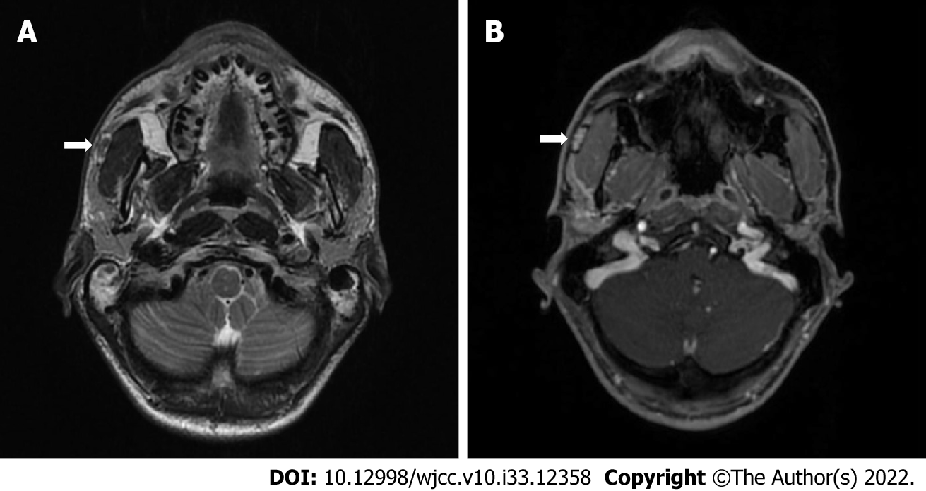
Figure 1 Magnetic resonance imaging T2-weighted image (A) and T1-weighted contrast-enhanced image.
A: An arrow shows an encapsulated tumor in the anterior portion of the right superficial lobe of the parotid gland (accessory parotid gland) with high-intensity signal capsule together with low-intensity signal core; B: The same tumor with the contrast accumulating core and no clear contrast accumulation in the capsule.
- Citation: Stankevicius D, Petroska D, Zaleckas L, Kutanovaite O. Hybrid intercalated duct lesion of the parotid: A case report. World J Clin Cases 2022; 10(33): 12358-12364
- URL: https://www.wjgnet.com/2307-8960/full/v10/i33/12358.htm
- DOI: https://dx.doi.org/10.12998/wjcc.v10.i33.12358


