Copyright
©The Author(s) 2022.
World J Clin Cases. Oct 6, 2022; 10(28): 10301-10309
Published online Oct 6, 2022. doi: 10.12998/wjcc.v10.i28.10301
Published online Oct 6, 2022. doi: 10.12998/wjcc.v10.i28.10301
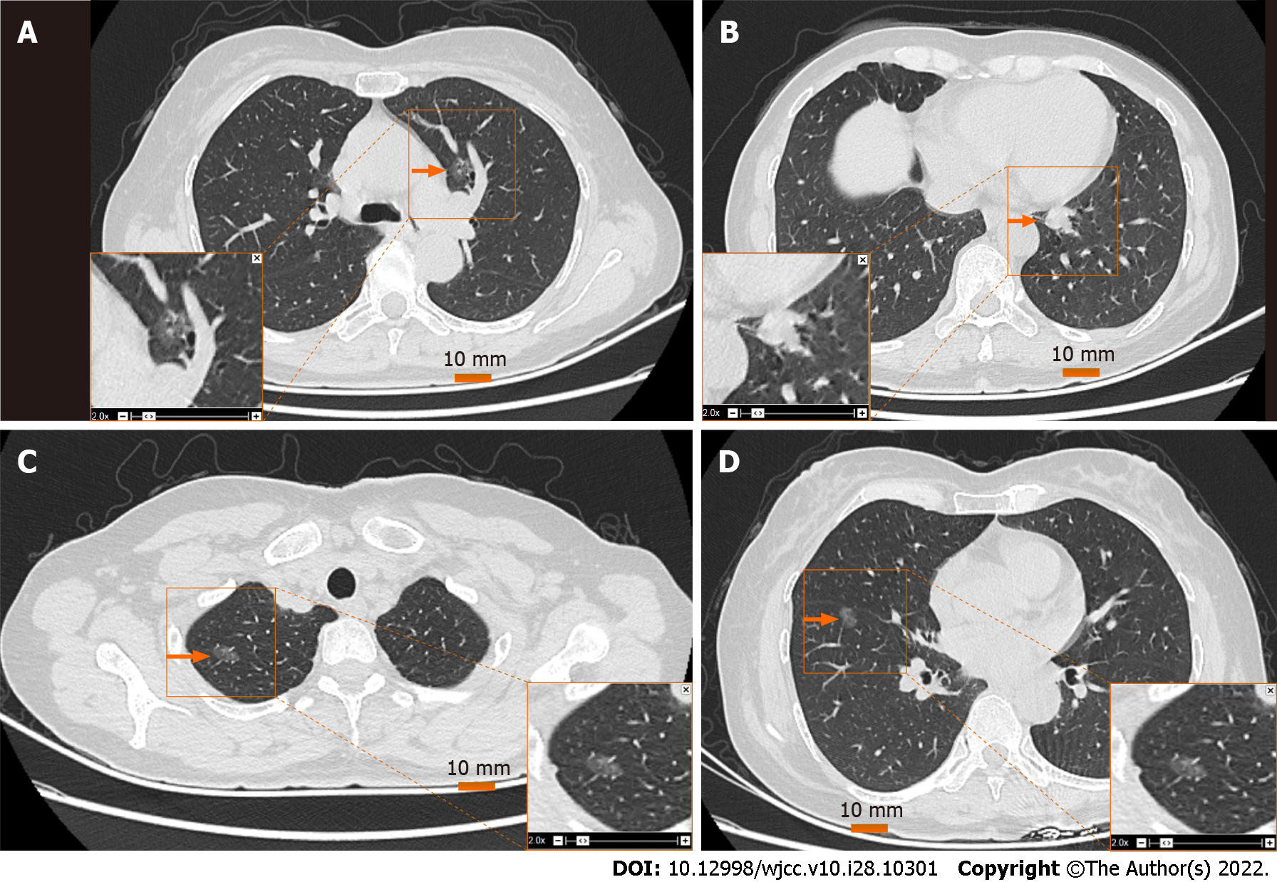
Figure 1 Computed tomography image of pulmonary nodules.
A: Left upper lung nodule (LS3), 11 mm in diameter; B: Left lower lung nodule (LS7), 20 mm × 14 mm × 10 mm; C: Upper right nodule (RS1), 8 mm in diameter; D: Lower right nodule (RS8), 8 mm in diameter. The orange arrow indicates the location of the nodule.
- Citation: Zhang DY, Liu J, Zhang Y, Ye JY, Hu S, Zhang WX, Yu DL, Wei YP. One-stage resection of four genotypes of bilateral multiple primary lung adenocarcinoma: A case report. World J Clin Cases 2022; 10(28): 10301-10309
- URL: https://www.wjgnet.com/2307-8960/full/v10/i28/10301.htm
- DOI: https://dx.doi.org/10.12998/wjcc.v10.i28.10301


