Published online Sep 6, 2022. doi: 10.12998/wjcc.v10.i25.9162
Peer-review started: April 27, 2022
First decision: June 16, 2022
Revised: June 28, 2022
Accepted: July 25, 2022
Article in press: July 25, 2022
Published online: September 6, 2022
Processing time: 121 Days and 7.6 Hours
Cholesterol crystal embolization (CCE) is a multisystemic and fatal disease with multiple clinical manifestations; however, there are few cases of idiopathic CCE. Here we report a patient with idiopathic CCE accompanied by atheroembolic renal disease and blue toes who had a relatively good prognosis in the short-term due to early treatment with corticosteroids and statins.
A 76-year-old man complained of coldness, numbness and purple color change in his left foot for 7 d. He had a feeling of fatigue, constipation, foamy urine, poor appetite and sleep. He had a lacunar infarction for 5 years and hypertension for 9 mo. Laboratory results showed elevated eosinophils, cholesterol, uric acid, serum creatinine, urea and 24 h urine analysis revealed proteinuria. A renal biopsy revealed atheroembolic renal disease. Taken together, these findings strongly supported the diagnosis of idiopathic CCE and atheroembolic renal disease.
Atheroembolic renal disease and blue toes syndrome can be caused by idiopathic CCE, and early treatment with corticosteroids is effective but requires further investigation.
Core Tip: Idiopathic cholesterol crystal embolization can induce atheroembolic renal disease and blue toes syndrome, and the early corticosteroids treatment is effective.
- Citation: Cheng DJ, Li L, Zheng XY, Tang SF. Idiopathic cholesterol crystal embolism with atheroembolic renal disease and blue toes syndrome: A case report. World J Clin Cases 2022; 10(25): 9162-9167
- URL: https://www.wjgnet.com/2307-8960/full/v10/i25/9162.htm
- DOI: https://dx.doi.org/10.12998/wjcc.v10.i25.9162
Cholesterol crystal embolization (CCE) is caused by diffusion of cholesterol atheroma debris and can be triggered by intraarterial interventions and anticoagulant therapy, leading to both ischemic and inflammatory damage to the target organ[1]. However, there are only a few reports of patients with idiopathic CCE who did not receive medication or arterial intervention[2,3]. As cholesterol atheroma can occlude all types of small arteries, the disease has various clinical manifestations involving organs such as the brain, eye, kidney, gastrointestinal system and skin, which makes CCE a fatal disease with poor prognosis[4]. In a review of 221 cases of histologically proven CCE, the mortality rate was as high as 80%[5]. Here we report a patient with idiopathic CCE accompanied by atheroembolic renal disease and blue toes who had a relatively good prognosis in the short-term due to early treatment with corticosteroids and statins.
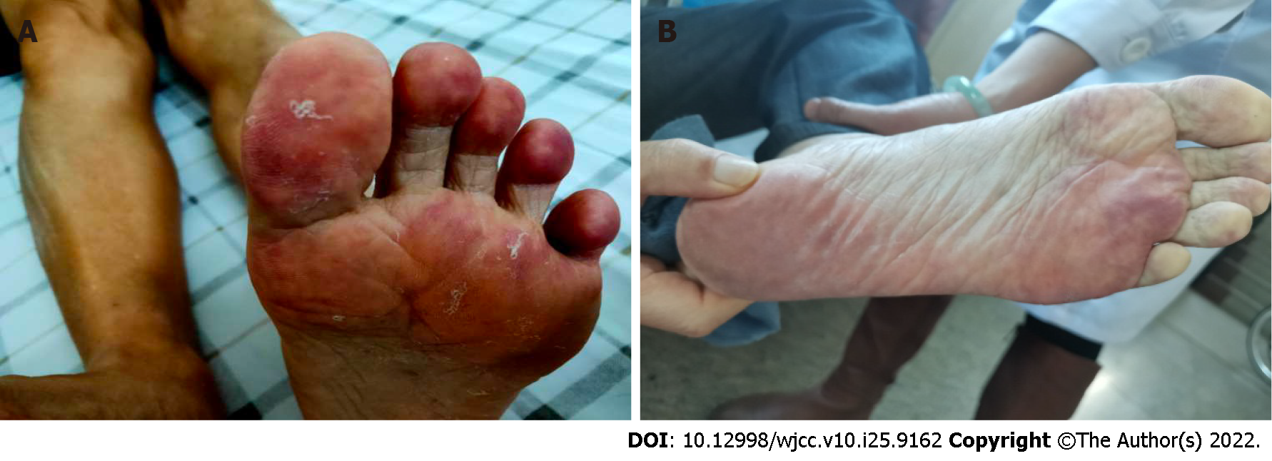
A 76-year-old man complained of coldness, numbness and purple color change (Figure 1A) in his left foot for 7 d. He had a feeling of fatigue, constipation, foamy urine, poor appetite and sleep, but no fever, headache or abdominal pain. His urine output was normal.
A 76-year-old man complained of coldness, numbness and purple color change in his left foot for 7 d. And he came to our hospital for treatments.
The patient had a lacunar infarction for five years and took three Fufang Xueshuantong capsules three times daily. He also had hypertension (maximum blood pressure: 189/102 mmHg) for 9 mo and took one tablet of amlodipine, atorvastatin calcium and fosinopril sodium tablets 10 mg once daily. There was no history of surgery.
Physical examination upon admission showed no abnormalities.
Laboratory findings (Table 1) showed elevated eosinophils, triglycerides, serum creatinine and urea. The 24 h urine analysis showed increased proteinuria.
| Items | Reference value | Results |
| Eosinophil count (x 109/L) | 0.05-0.3 | 3.46 |
| Serum creatinine (mmol/L) | 57-97 | 239 |
| Serum urea (mmol/L) | 3.6-9.5 | 23.32 |
| Cholesterol (mmol/L) | 2.6-5.2 | 3.90 |
| Triglycerides (mmol/L) | 0.34-1.60 | 1.63 |
| HDL-C (mmol/L) | > 1.04 | 1.43 |
| LDL-C (mmol/L) | ≤ 3.37 | 2.19 |
| Urinary protein (g/24 h) | ≤ 0.15 | 0.89 |
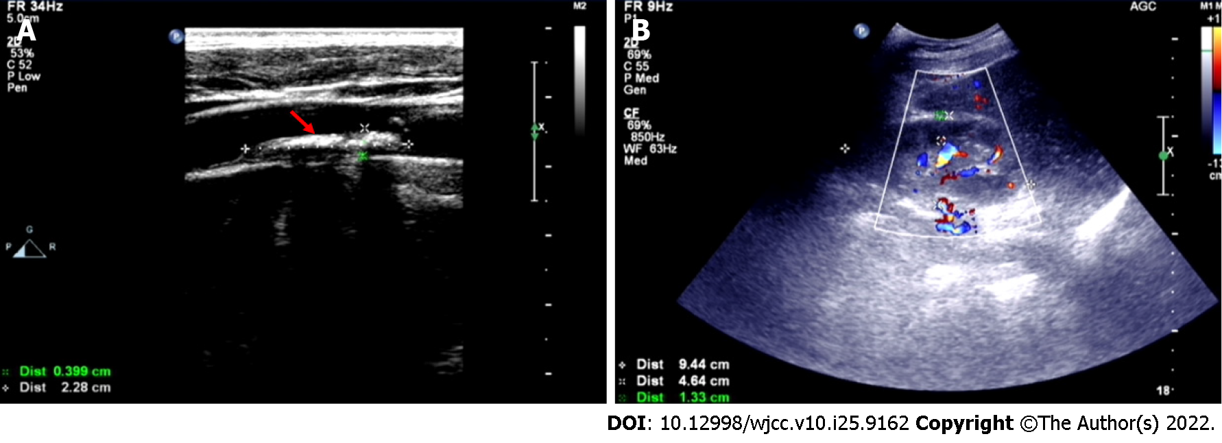
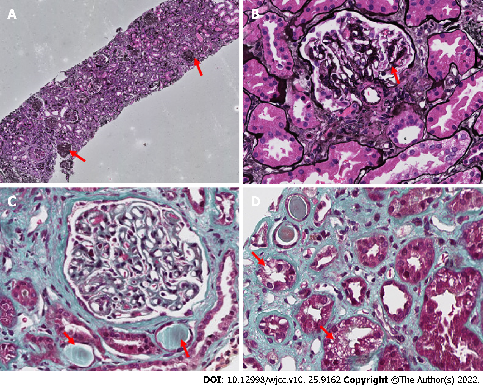
Doppler ultrasonography showed carotid atherosclerosis with carotid plaque formation (Figure 2A) and both kidneys were of normal size; however, kidney parenchymal thickness (approximately 13 mm) was slightly thinner (Figure 2B). Liver, spleen and pancreas were normal. A renal biopsy (Figure 3) showed glomerulus ischemic globular sclerosis in all 41 glomerulus; and mesangial cells and stroma showed slight hyperplasia in the rest of the glomerulus, one small cell fibrous crescent, and some of the glomerular capillaries were ischemic and wrinkled. With regard to the renal tubules, epithelial cells showed cavitation and granular degeneration, crystal nucleation within some renal tubules, a few renal tubules demonstrated cavity expansion, disappearance of brush border, multifocal and atrophy (shrinkage area of approximately 60%); diffuse and patchy infiltration of the kidney interstitium by lymphocytes, plasma cells and a few eosinophils; small artery wall thickening with a narrow lumen, endometrial hyperplasia, and cholesterol embolism filling several small arteries. Congo and oxidized Congo red were negative. Immunofluorescence showed no deposition of immune complexes.
The patient was diagnosed with idiopathic cholesterol crystal embolization and atheroembolic renal disease.
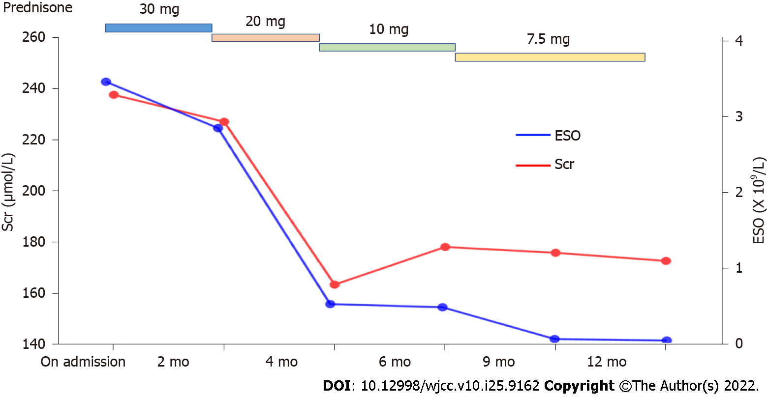
He was treated with 30 mg of prednisone acetate tablets once daily during hospitalization and the dosage was gradually tapered after discharge. The patient also received a 20 mg tablet of atorvastatin calcium, 50 mg tablet of clopidogrel bisulfate, 10 mg tablet of fosinopril sodium and 10 mg of amlodipine once a day, respectively.
During the 12-mo follow-up period, the color of his left foot gradually returned to normal (Figure 1B). The gradual reduction in prednisone acetate is shown in Figure 4. Following treatment with prednisone, eosinophils and serum creatine gradually decreased after discharge (Figure 4).
CCE is a multisystemic disease with various clinical manifestations induced by atherosclerotic plaques, and these plaques are composed of platelets, fibrin, necrotic cell debris, and cholesterol crystals (CCs)[6]. Hemodynamic changes, intraplaque hemorrhage, and inflammation, which may occur spontaneously (namely idiopathic CCE) or due to invasive procedures, can induce plaque erosion and rupture that expose the plaque components to the systemic circulation. Initially, CCs only cause ischemic injury; but the subsequent inflammatory reaction aggravates end-organ injury. Endothelial injury, oxidative stress, activation of the renin-angiotensin-aldosterone system, leukocyte aggregation, complement activation, and release of leukocyte enzymes are all considered responsible for end-organ injury[1,7]. Approximately 80% of cases of CCE have been reported to be due to anticoagulation therapy and catheter manipulation, and there are few cases of idiopathic CCE[8]. The patient with definite path
With regard to treatment, secondary prevention of cardiovascular disease is of utmost importance in these patients, such as statins, antiplatelet therapy, cessation of smoking, and control of blood pressure, weight, and glycemia. Furthermore, anti-inflammatory treatment such as corticosteroids and cyclophosphamide are alternative choices but these drugs have not been evaluated in randomized controlled trials[7]. Some studies demonstrated improved renal function with high-dose corticosteroid treatment in patients with CCE[10,11]. One patient with leg ulceration caused by CCE was reported to improve with colchicine and corticosteroids[12]. However, several studies have shown that corticosteroid therapy results in good renal outcome in CCE patients in the short-term, but does not have a favorable effect on long-term renal outcome[13]. Statins, prednisone, clopidogrel and antihypertensive agents were administered to our patient. In the 12 mo follow-up period, his renal function gradually improved and the level of eosinophils gradually decreased, which demonstrated that prednisone treatment was effective. However, the long-term effect of corticosteroid treatment and the dose reduction regimen require further investigation in randomized controlled trials.
Atheroembolic renal disease and blue toes syndrome can be caused by idiopathic CCE, and early treatment with corticosteroids is effective but requires further study.
Provenance and peer review: Unsolicited article; Externally peer reviewed.
Peer-review model: Single blind
Specialty type: Urology and nephrology
Country/Territory of origin: China
Peer-review report’s scientific quality classification
Grade A (Excellent): 0
Grade B (Very good): 0
Grade C (Good): C, C, C
Grade D (Fair): 0
Grade E (Poor): 0
P-Reviewer: Corso G, Italy; Esposito P, Italy; Moreno-Gómez-Toledano R, Spain S-Editor: Ma YJ L-Editor: A P-Editor: Ma YJ
| 1. | Saric M, Kronzon I. Cholesterol embolization syndrome. Curr Opin Cardiol. 2011;26:472-479. [RCA] [PubMed] [DOI] [Full Text] [Cited by in Crossref: 35] [Cited by in RCA: 33] [Article Influence: 2.4] [Reference Citation Analysis (0)] |
| 2. | Higo S, Hirama A, Ueda K, Mii A, Kaneko T, Utsumi K, Iino Y, Katayama Y. A patient with idiopathic cholesterol crystal embolization: effectiveness of early detection and treatment. J Nippon Med Sch. 2011;78:252-256. [RCA] [PubMed] [DOI] [Full Text] [Cited by in Crossref: 4] [Cited by in RCA: 4] [Article Influence: 0.3] [Reference Citation Analysis (0)] |
| 3. | Tashiro M, Ito K, Saito T. An autopsy case of idiopathic cholesterol embolism. Clin Exp Nephrol. 2013;17:142-143. [RCA] [PubMed] [DOI] [Full Text] [Cited by in Crossref: 2] [Cited by in RCA: 2] [Article Influence: 0.2] [Reference Citation Analysis (0)] |
| 4. | Kuwatani M, Sakamoto N. Angiogenic Dreaded Killer: Cholesterol Crystal Embolization. Intern Med. 2021;60:825-826. [RCA] [PubMed] [DOI] [Full Text] [Full Text (PDF)] [Cited by in Crossref: 1] [Cited by in RCA: 1] [Article Influence: 0.3] [Reference Citation Analysis (0)] |
| 5. | Fine MJ, Kapoor W, Falanga V. Cholesterol crystal embolization: a review of 221 cases in the English literature. Angiology. 1987;38:769-784. [RCA] [PubMed] [DOI] [Full Text] [Cited by in Crossref: 296] [Cited by in RCA: 258] [Article Influence: 6.8] [Reference Citation Analysis (0)] |
| 6. | Kronzon I, Saric M. Cholesterol embolization syndrome. Circulation. 2010;122:631-641. [RCA] [PubMed] [DOI] [Full Text] [Cited by in Crossref: 108] [Cited by in RCA: 114] [Article Influence: 7.6] [Reference Citation Analysis (0)] |
| 7. | Ozkok A. Cholesterol-embolization syndrome: current perspectives. Vasc Health Risk Manag. 2019;15:209-220. [RCA] [PubMed] [DOI] [Full Text] [Full Text (PDF)] [Cited by in Crossref: 21] [Cited by in RCA: 43] [Article Influence: 7.2] [Reference Citation Analysis (0)] |
| 8. | Scolari F, Tardanico R, Zani R, Pola A, Viola BF, Movilli E, Maiorca R. Cholesterol crystal embolism: A recognizable cause of renal disease. Am J Kidney Dis. 2000;36:1089-1109. [RCA] [PubMed] [DOI] [Full Text] [Cited by in Crossref: 194] [Cited by in RCA: 162] [Article Influence: 6.5] [Reference Citation Analysis (0)] |
| 9. | Tanaka H, Yamana H, Matsui H, Fushimi K, Yasunaga H. Proportion and risk factors of cholesterol crystal embolization after cardiovascular procedures: a retrospective national database study. Heart Vessels. 2020;35:1250-1255. [RCA] [PubMed] [DOI] [Full Text] [Cited by in Crossref: 4] [Cited by in RCA: 4] [Article Influence: 0.8] [Reference Citation Analysis (0)] |
| 10. | Fabbian F, Catalano C, Lambertini D, Bordin V, Di Landro D. A possible role of corticosteroids in cholesterol crystal embolization. Nephron. 1999;83:189-190. [RCA] [PubMed] [DOI] [Full Text] [Cited by in Crossref: 33] [Cited by in RCA: 33] [Article Influence: 1.3] [Reference Citation Analysis (0)] |
| 11. | Desai M, Ram R, Prayaga A, Dakshinamurty KV. Cholesterol crystal embolization (CCE): Improvement of renal function with high-dose corticosteroid treatment. Saudi J Kidney Dis Transpl. 2011;22:327-330. [RCA] [PubMed] [DOI] [Full Text] [Cited by in Crossref: 4] [Cited by in RCA: 2] [Article Influence: 0.3] [Reference Citation Analysis (0)] |
| 12. | Verneuil L, Ze Bekolo R, Dompmartin A, Comoz F, Marcelli C, Leroy D. Efficiency of colchicine and corticosteroids in a leg ulceration with cholesterol embolism in a woman with rheumatoid arthritis. Rheumatology (Oxford). 2003;42:1014-1016. [RCA] [PubMed] [DOI] [Full Text] [Cited by in Crossref: 3] [Cited by in RCA: 4] [Article Influence: 0.2] [Reference Citation Analysis (0)] |
| 13. | Nakayama M, Izumaru K, Nagata M, Ikeda H, Nishida K, Hasegawa E, Ohta Y, Tsuchihashi T, Urabe K. The effect of low-dose corticosteroids on short- and long-term renal outcome in patients with cholesterol crystal embolism. Ren Fail. 2011;33:298-306. [RCA] [PubMed] [DOI] [Full Text] [Cited by in Crossref: 20] [Cited by in RCA: 23] [Article Influence: 1.6] [Reference Citation Analysis (0)] |


