Published online Sep 6, 2022. doi: 10.12998/wjcc.v10.i25.9078
Peer-review started: April 11, 2022
First decision: June 16, 2022
Revised: June 28, 2022
Accepted: July 25, 2022
Article in press: July 25, 2022
Published online: September 6, 2022
Processing time: 137 Days and 6.3 Hours
Canines are the most important teeth in the dentition. Usually, doctors choose to remove premolars rather than canines. Canine extraction is extremely rare in orthodontic treatment. However, dentists sometimes encounter situations in which canines require extraction due to defects caused by improper medical treatment.
The present study reports a case of a class II adult patient treated with the extraction of maxillary canines and right mandibular second premolar. After postactive treatment for 28 mo, then the canines were substituted by the upper first premolar, a satisfactory occlusal was established, the lips were competent, and the profile was improved. Intraoral pictures and X-ray data retrieved 3 years after the end of orthodontic treatment demonstrated the possibility of canine extraction and premolar substitution of canines in function and beauty.
The extraction of canines and substitution by first premolars could be a feasible orthodontic treatment.
Core Tip: A modified orthodontic treatment substitution of canines by first premolars, Unconventional orthodontic treatment programs eliminated the risk factors and uncertain results. It has very practical clinical significance.
- Citation: Li FF, Li M, Li M, Yang X. Modified orthodontic treatment of substitution of canines by first premolars: A case report. World J Clin Cases 2022; 10(25): 9078-9086
- URL: https://www.wjgnet.com/2307-8960/full/v10/i25/9078.htm
- DOI: https://dx.doi.org/10.12998/wjcc.v10.i25.9078
Since canines are the longest teeth in the oral cavity, they play a critical role in occlusal function and aesthetics[1,2]. The smooth shape gives the canines a good self-cleaning effect with less severe caries. Due to the large root surface areas and improved crown to root ratio, a high occlusal force can be supported[3]. Canines are not injured easily because of their position at the middle corner of the dental arch. The canines guide the teeth into the intercuspal position[4], which in turn supports the corners of the mouth, maintaining the smile and facial aesthetics[5]. For these reasons, canine extraction is uncommon in modern orthodontic treatment. However, the abnormal development of canines is not uncommon. The prevalence of ectopic impact was 1.38%, canine agenesis was 0.06% and ectopic canine was 5.5%[6]. The present study reports a case of an adult with class II occlusion and lip protrusion who had crowding maxillary canines and crowns removed artificially many years ago due to unaesthetic looks. Dentists encounter situations wherein canine extraction seems inevitable. We have to make a choice between abandoning or retaining the canine tooth. After treatment, a satisfactory occlusal correlation with an aesthetic smile was achieved by extracting the severely damaged canines and closing the extraction space. This avoided the complicated course of treatment, high cost, and follow-up risk of the canine restoration approach.
A 20-year-old male complained of an irregular teeth arrangement.
The patient is not satisfied with his teeth recently, which affects his social interaction. Desire for improvement of teeth aesthetics.
Bilateral maxillary canine crowns were removed many years ago by inappropriate treatment.
No similar family history was reported.
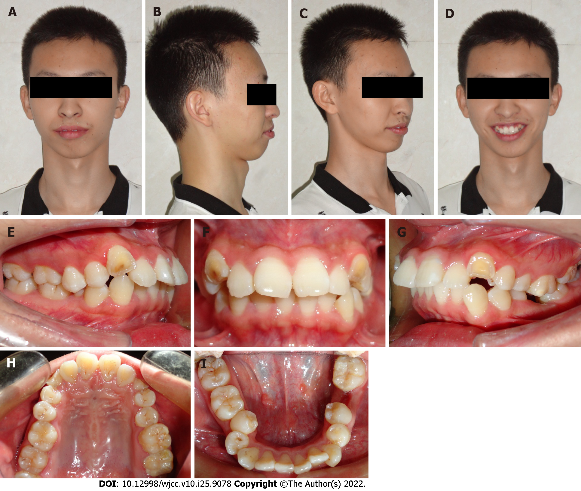
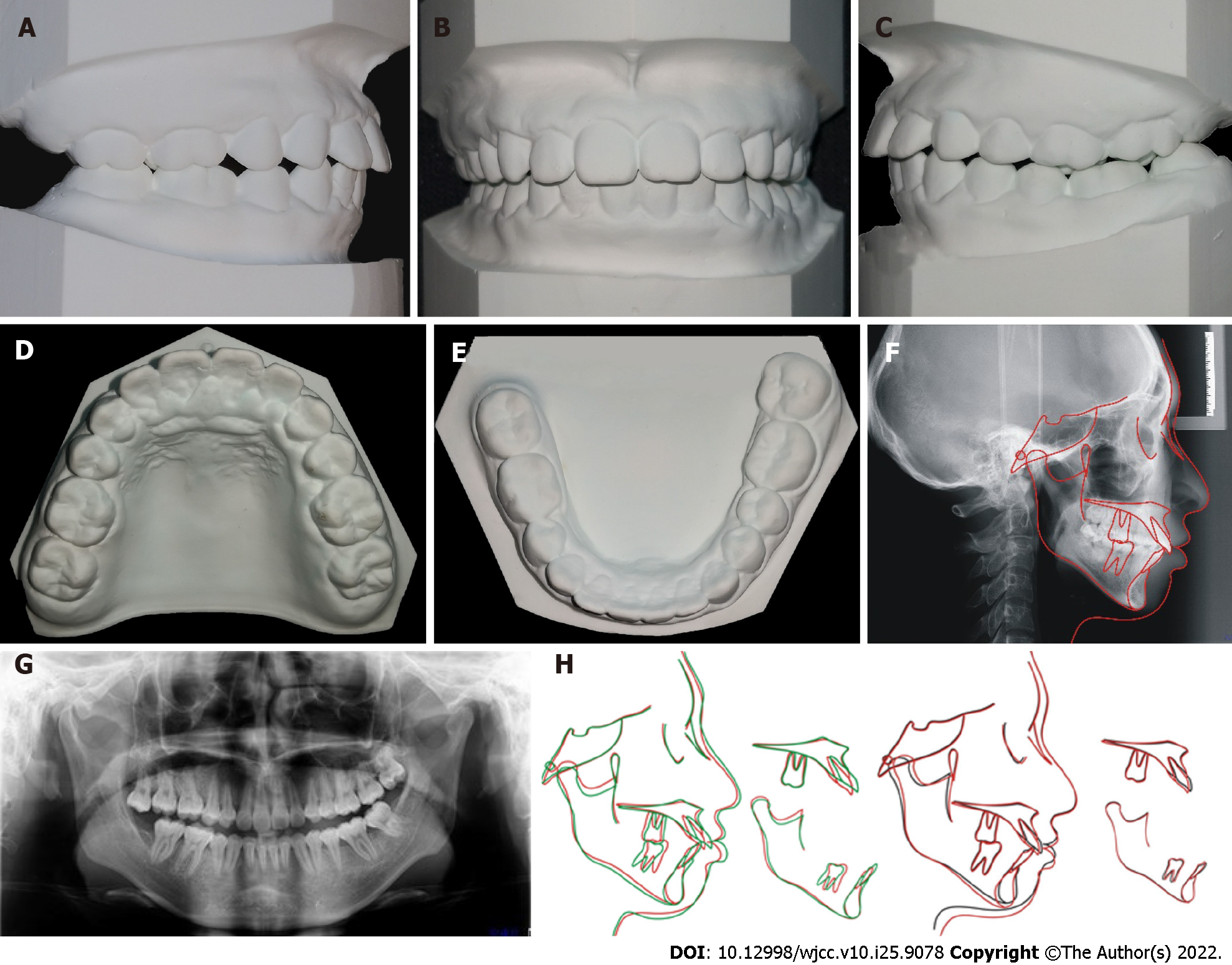
The facial analysis showed facial asymmetry; the left side was larger than the right side. Additionally, left deviation of the chin, labial protrusion, and lip tension was observed. The intraoral examination showed moderate crowding in the maxillary and mandibular arches; bilateral maxillary canines were ectopic, and crowns were removed previously by a barbaric treatment without any root canal therapy because of poor aesthetics. The upper and lower anterior teeth were inclined, the lower middle line deviated approximately 2 mm to the left, the mandibular first molar was missing while the maxillary first molar was overerupted, and a crossbite of the right mandibular second premolar was also noticeable (Figure 1). A Class I molar relationship was present on the right side.
Normal.
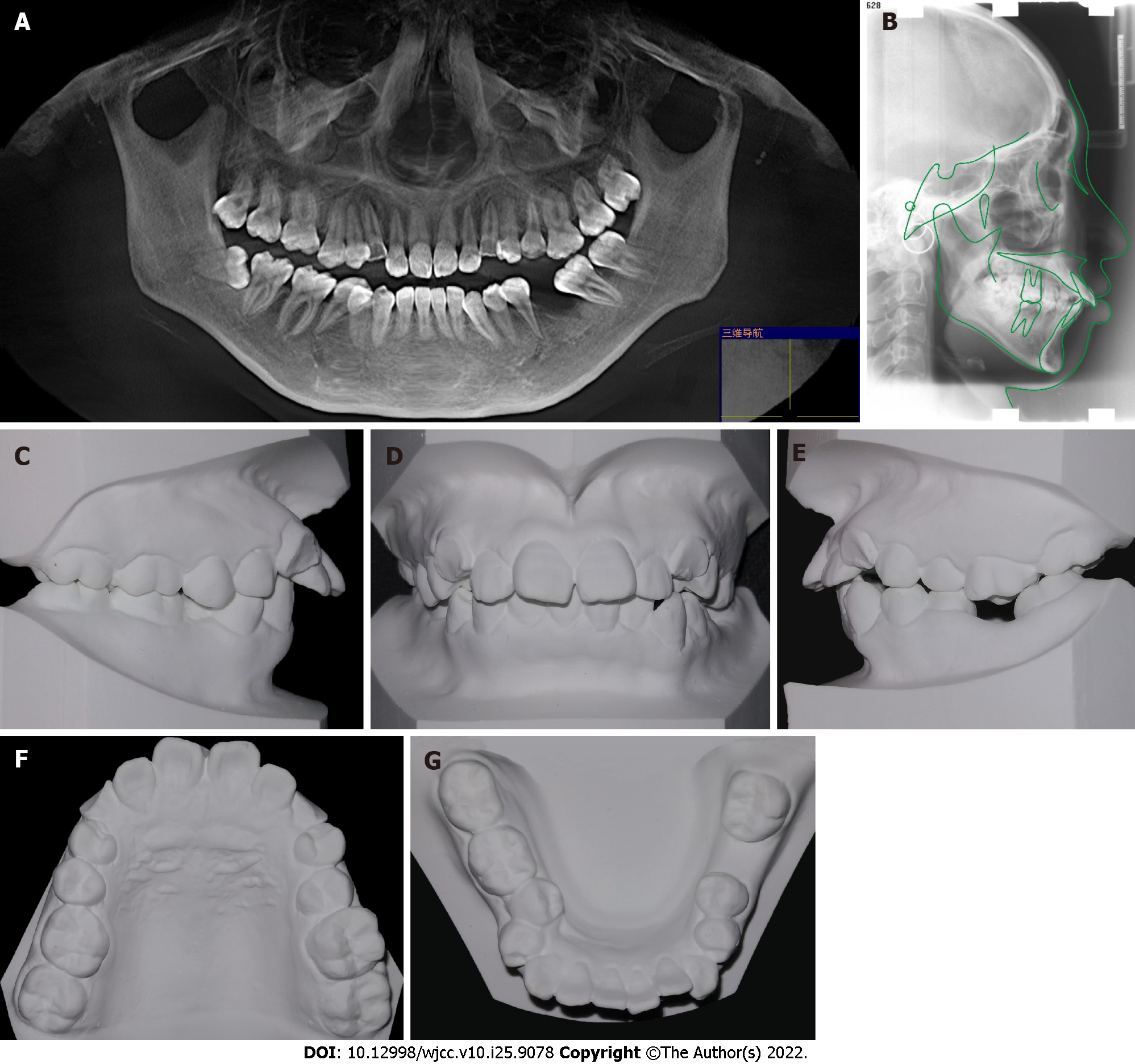
The panoramic radiograph showed periapical inflammation of the bilateral maxillary canines (Figure 2A). The cephalometric analysis showed a high angle and class II skeletal correlation because of maxillary overgrowth (Figure 2B-G, Table 1). The patient did not state a similar family history.
| Measurement | Pretreatment | Post-treatment |
| SNA (°) | 88.5 | 88.0 |
| SNB (°) | 81.7 | 82.8 |
| ANB (°) | 5.8 | 5.2 |
| U1-NA (mm) | 8 | 5 |
| U1-NA (°) | 35.9 | 26.2 |
| L1-NB (mm) | 9 | 8 |
| L1-NB (°) | 30.7 | 31.7 |
| MP-FH (°) | 29 | 30 |
| Y (°) | 61 | 61 |
| U1-SN (°) | 56.5 | 61 |
| L1-MP (°) | 97 | 98 |
| U1-L1 (°) | 107.5 | 115.3 |
Angle class II and skeletal class II high Angle cases; Lip protrusion; moderate crowding; deep overjet and overbite; tooth midline inconsistency; right second premolar positive crossbite.
The treatment objectives were to maintain a neutral occlusion on the right and distal occlusal relationship on the left, align the upper and lower occlusal arches, solve the aesthetic problems of the upper canines, and retract the inclined anterior teeth, improve facial appearance, and reduce lip protrusion.
Restoration and extraction of maxillary canines were considered. Prosthodontic doctors recommend prosthetic replacement for maxillary canines after traditional root canal therapy; however, the prognosis is not optimistic. The upper first premolars still need to be extracted. In this patient, crowding was associated with dentoalveolar protrusion, which was efficiently corrected by maxillary first premolar extractions. Subsequently, lip incompetence is addressed, and space is created for easy alignment of crowding canines. Nonetheless, the patient will bear high restoration costs, and the treatment process will be much more complicated. Additionally, defective fixed prostheses may contribute to the progression of periodontal diseases[7].
Right mandibular second premolar extractions would resolve crossbite, reduce dentoalveolar protrusion, and improve lip competence. Furthermore, mandibular right second premolar extractions (compared to first premolar extractions) might reduce the amount of retraction during the gap closure, thereby reducing the flattening of the profile and providing sufficient space for the eruption of molars.
To simplifying the treatment process, reducing the cost of restoration, and taking into account the patient’s wishes, the final plan contained surgical extraction of the upper canines and the mandibular second premolars; the maxillary first premolars were planned to substitute the canines. This approach would reduce the time of treatment, reduce dentoalveolar protrusion, and improve facial appearance.
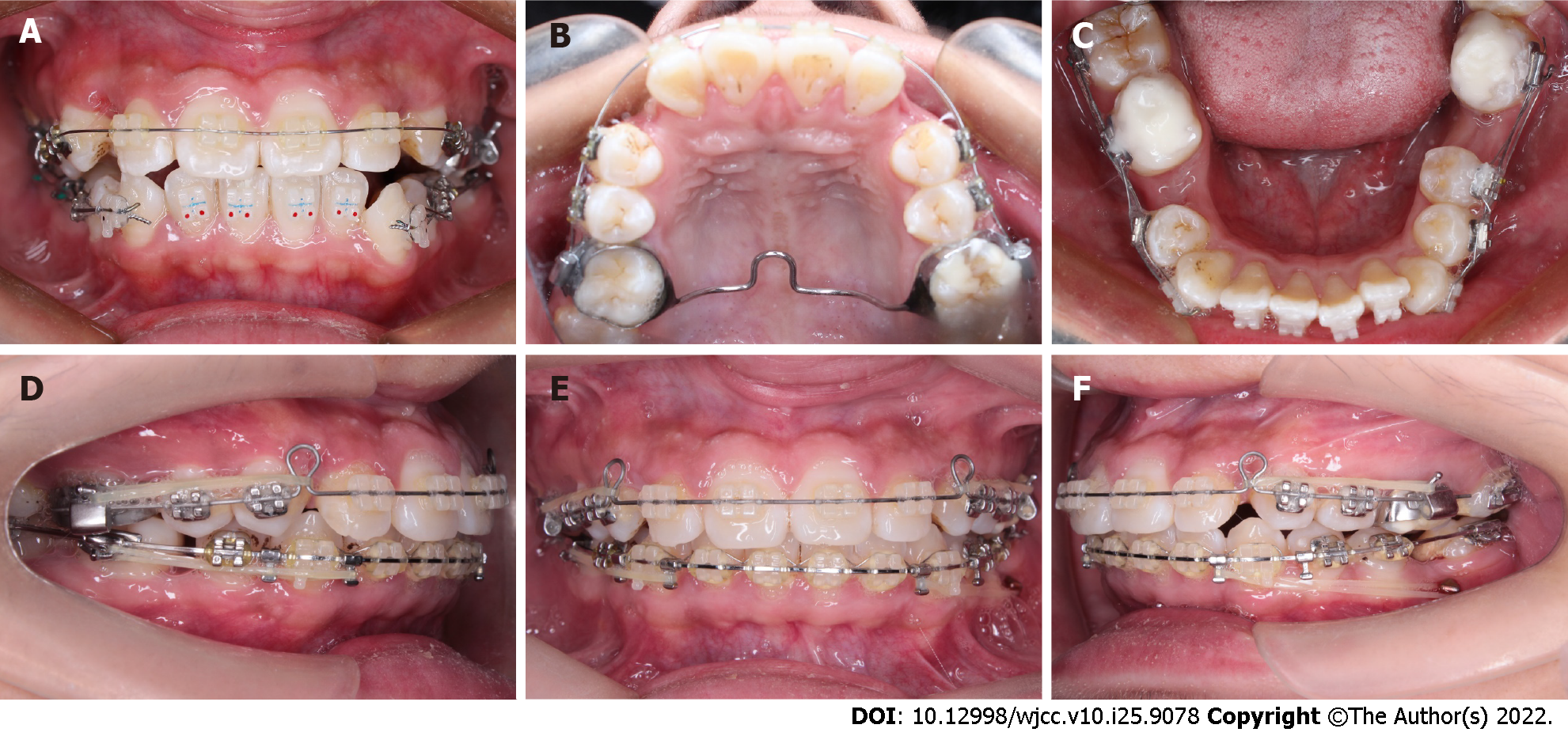
After tooth extraction according to the plan, fixed appliances were bonded. At the beginning of the treatment, a transpalatal arch was used to control the vertical height of the posterior teeth (Figure 3A-C). Because the crown on the mandibular canine is inclined toward the mesial, to avoid inclination of the lower anterior teeth when aligning, the fragment arch technique was used to retract the mandibular canine first, while the lower incisor was temporarily not included in the treatment system A 0.014/0.016/0.018-inch nickel-titanium wire was used to align the upper and lower dental arches. Leveling the maxillary and mandibular arches with 0.019 × 0.025-inch nickel-titanium wires. In the following 18 mo, 0.019 × 0.025-inch stainless steel rectangular wires were replaced in both arches to close the extraction spaces. The left mandibular space was posterior to the arch, and hence, a miniscrew was used to strengthen the anchorage and retract the lower anterior teeth to reduce the protrusion. After 20 mo of orthodontic treatment, the anterior maxillary teeth were still slightly inclined. Next, we replaced the 0.018-inch stainless steel wire to further reduce the inclination (Figure 3D-F).
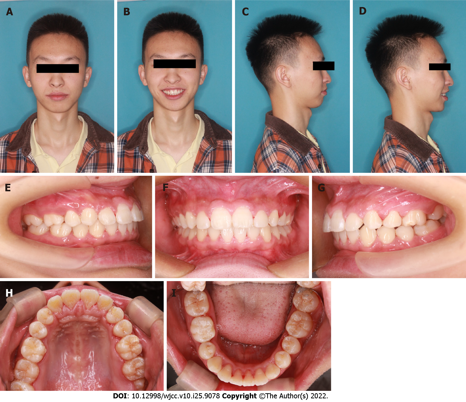
After treatment, the patient’s dentition and facial protrusion were greatly improved, the mandibular deviation was alleviated, the left and right sides were symmetrical, the overlap was well covered, and the occlusion relationship was satisfactory. However, the lower anterior teeth showed slight lip inclination, and X-ray analysis did not show any significant root resorption (Figure 5G) at 2.5 years post-correction. The vertical height was effectively controlled during the treatment. A significant clockwise rotation of the mandible did not occur. The sagittal anchorage of the posterior teeth was well retained. The lower anterior teeth were depressed, the anterior teeth were retracted, and the lip protrusion was significantly improved (Figures 4 and 5). After treatment, the dentition was orderly, the occlusion was satisfactory, the masticatory function was normal, and the natural teeth were preserved without crown restoration or implant therapy; this treatment process was simplified in a cost-efficient manner. Subsequently, the profile and smile improved obviously, and no face collapse was detected after the extraction of canines as expected at the beginning of the treatment.
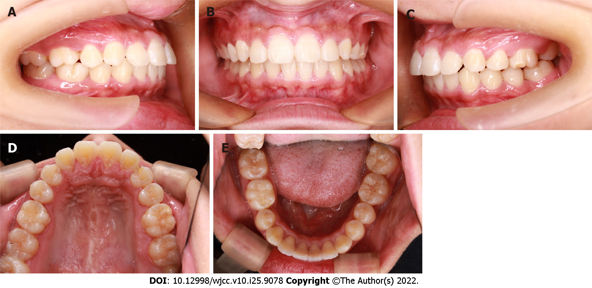
Hawley retainers were delivered to the patient. Intraoral images were taken 3 years after the end of fixed appliance treatment (Figure 6). The arch form and dental alignment were adequate. The patient still had an appropriate occlusion. No temporomandibular joint or muscle problems reported during the treatment and maintenance period. The temporomandibular joints did not show any pain or clicking on opening and closing. No deviation upon opening of the jaws was found. No premature contacts were observed.
Canines are one of the key elements in the maxillofacial system. Considering the smile aesthetics, occlusal function, and temporomandibular joint health, canine extraction is relatively rare in modern orthodontic treatment. Occasionally, the canines have to be extracted due to severe ectopic eruption, congenital developmental defects, a large area of caries, and congenital loss of canines. The present case was an adult male. According to the facial protrusion view, the four first premolars needed to be extracted; however, the oral condition of the patient was complicated, the maxillary canines had been severely damaged for many years, and the X-ray showed chronic root apical inflammation. Prosthetic replacement of the maxillary canine after perfect root canal therapy is the primary repair method. Intriguingly, utilizing a core crown can result in several complications. In addition, it was found that endodontically treated teeth moved as readily as vital teeth, albeit with a large frequency of root resorption in the endodontically treated teeth due to the presence of apical deltas, the material used, and many other factors[8]. Teeth undergoing root canal treatment might be predisposed to progressive severe inflammatory root resorption during orthodontic tooth movement[9]. Some scholars point out that if the patient has a past history of trauma or root canal treatment, radiographic monitoring should be carried out[7]. Some research emphasizes the importance of orthodontic treatment planning, following conventional treatment protocols and proper application of orthodontic forces to prevent root resorption and achieve successful outcomes[10]. Furthermore, it might be difficult to achieve canine-guided occlusion by crown restoration. A similar issue was detected with canine extraction. According to some studies, canine guidance could be partially supplied by premolar guidance or group function, and substitution of the first premolars for the canines is effective in attaining a functional occlusion[11]. Reportedly, 81% of the natural dentition had the function of group occlusion, but only 5% of the natural dentition had canines guiding the occlusion[12]. The long-term stability of this canine guidance established by orthodontic treatment has yet to be discussed[13]. Thus, the establishment of group function is more practical than canine guidance in orthodontic treatment. There are many ways to optimize functionality in canine extraction cases, and individualized extrusion, mesial rotation, and crown lingual torque of the upper first premolars were considered to eliminate occlusal interference and imitate canine prominence[11]. In this case, the health of the temporomandibular joint is also one of the considerations. Interestingly, the patient did not have discomfort in the temporomandibular joint during or after the treatment. Rosa et al[14] reported that patients with premolars replacing the canines did not exhibit joint problems in long-term observation. Second, the treatment progress is much more complicated by the orthodontic restoration approach. The canines need to be distally retracted, but the fixed appliance does not bond strongly to the residual canines, necessitating the placement of bands on the maxillary canines, which presents a risk of periodontal problems[15]. Another issue related to crown restoration problems is smile aesthetics. Canines are essential for aesthetics, giving the patient correct dental and gingival symmetry[1]. The canine eminence provides support to the nasolabial fold, which will help the patient have better facial aging, and a greater sense of width in the smile is achieved. In this patient, maxillary canine gingival margins were slightly recessed because of long-term crown defects. Some investigators suggested that premolars can replace canines in facial aesthetics, and no significant difference was detected in facial smile aesthetics between the general population and professional physicians[16]. Obviously, surgical extraction of the upper canines reduced the risk factors and uncertain results.
Next, we planned to extract the right lower second premolar because it had a crossbite. However, it was difficult to relieve crowding of the lower anterior teeth. At the beginning of treatment, the segment arch was used to move the mandibular canines distally. A microimplant on the lower right side was employed to retract the mandibular anterior teeth; however, according to the posttreatment cephalometrics, the lower anterior teeth showed a slight inclination at the end of the treatment. This could be related to the class II skeletal type. However, at 3 years after posttreatment, we also observed that the inclination of the maxillary incisor had slightly relapsed. The long-term functionality of a premolar in place of a canine has yet to be determined.
In this case, canine extraction avoids root canal treatment and crown restoration. Natural teeth are preserved, and the treatment is simplified. On the left side, we tracked the mesial second and third molars and avoided a dental implant at 36. For similar cases, the extraction of canines and substitution by first premolars are highly recommended for eliminating the risk factors and uncertain results.
Provenance and peer review: Unsolicited article; Externally peer reviewed.
Peer-review model: Single blind
Specialty type: Dentistry, oral surgery and medicine
Country/Territory of origin: China
Peer-review report’s scientific quality classification
Grade A (Excellent): 0
Grade B (Very good): 0
Grade C (Good): C, C
Grade D (Fair): 0
Grade E (Poor): 0
P-Reviewer: Heboyan A, Armenia; Kukiattrakoon B, Thailand S-Editor: Chen YL L-Editor: A P-Editor: Wu RR
| 1. | Lee MY, Park JH, Jung JG, Chae JM. Forced eruption of a palatally impacted and transposed canine with a temporary skeletal anchorage device. Am J Orthod Dentofacial Orthop. 2017;151:1148-1158. [RCA] [PubMed] [DOI] [Full Text] [Cited by in Crossref: 6] [Cited by in RCA: 6] [Article Influence: 0.8] [Reference Citation Analysis (0)] |
| 2. | Lorente C, Lorente P, Perez-Vela M, Esquinas C, Lorente T. Orthodontic management of a complete and an incomplete maxillary canine-first premolar transposition. Angle Orthod. 2020;90:457-466. [RCA] [PubMed] [DOI] [Full Text] [Cited by in Crossref: 9] [Cited by in RCA: 5] [Article Influence: 1.0] [Reference Citation Analysis (0)] |
| 3. | Clark JR, Evans RD. Functional occlusion: I. A review. J Orthod. 2001;28:76-81. [RCA] [PubMed] [DOI] [Full Text] [Cited by in Crossref: 54] [Cited by in RCA: 40] [Article Influence: 1.7] [Reference Citation Analysis (0)] |
| 4. | Thornton LJ. Anterior guidance: group function/canine guidance. A literature review. J Prosthet Dent. 1990;64:479-482. [RCA] [PubMed] [DOI] [Full Text] [Cited by in Crossref: 49] [Cited by in RCA: 38] [Article Influence: 1.1] [Reference Citation Analysis (0)] |
| 5. | Mirabella D, Giunta G, Lombardo L. Substitution of impacted canines by maxillary first premolars: a valid alternative to traditional orthodontic treatment. Am J Orthod Dentofacial Orthop. 2013;143:125-133. [RCA] [PubMed] [DOI] [Full Text] [Cited by in Crossref: 9] [Cited by in RCA: 5] [Article Influence: 0.4] [Reference Citation Analysis (0)] |
| 6. | Jain S, Debbarma S. Patterns and prevalence of canine anomalies in orthodontic patients. Med Pharm Rep. 2019;92:72-78. [RCA] [PubMed] [DOI] [Full Text] [Full Text (PDF)] [Cited by in Crossref: 6] [Cited by in RCA: 10] [Article Influence: 1.7] [Reference Citation Analysis (0)] |
| 7. | Heboyan A, Avetisyan A, Margaryan M, Manrikyan M, Vardanyan I. Tooth Root Resorption Conditioned by Orthodontic Treatment. Oral Hlth and Dent Sci. 2019;3:1-8. [DOI] [Full Text] |
| 8. | Consolaro A, Consolaro RB. Orthodontic movement of endodontically treated teeth. Dental Press J Orthod. 2013;18:2-7. [RCA] [PubMed] [DOI] [Full Text] [Cited by in Crossref: 7] [Cited by in RCA: 8] [Article Influence: 0.7] [Reference Citation Analysis (0)] |
| 9. | Esteves T, Ramos AL, Pereira CM, Hidalgo MM. Orthodontic root resorption of endodontically treated teeth. J Endod. 2007;33:119-122. [RCA] [PubMed] [DOI] [Full Text] [Cited by in Crossref: 36] [Cited by in RCA: 35] [Article Influence: 1.9] [Reference Citation Analysis (0)] |
| 10. | Heboyan A. Clinical Case of Root Resorption Due to Improper Orthodontic Treatment. J of Res in Medi and Dent Sci. 2019;7:91-93. |
| 11. | Sumiyoshi K, Ishihara Y, Komori H, Yamashiro T, Kamioka H. Orthodontic Treatment of a Patient with Bilateral Congenitally Missing Maxillary Canines: The Effects of First Premolar Substitution on the Functional Outcome. Acta Med Okayama. 2016;70:57-62. [RCA] [PubMed] [DOI] [Full Text] [Cited by in RCA: 2] [Reference Citation Analysis (2)] |
| 12. | Weinberg LA. The prevalence of tooth contact in eccentric movements of the jaw: its clinical implications. J Am Dent Assoc. 1961;62:402-406. [RCA] [PubMed] [DOI] [Full Text] [Cited by in Crossref: 13] [Cited by in RCA: 13] [Article Influence: 0.2] [Reference Citation Analysis (0)] |
| 13. | Rinchuse DJ, Kandasamy S, Sciote J. A contemporary and evidence-based view of canine protected occlusion. Am J Orthod Dentofacial Orthop. 2007;132:90-102. [RCA] [PubMed] [DOI] [Full Text] [Cited by in Crossref: 64] [Cited by in RCA: 52] [Article Influence: 2.9] [Reference Citation Analysis (0)] |
| 14. | Rosa M, Lucchi P, Ferrari S, Zachrisson BU, Caprioglio A. Congenitally missing maxillary lateral incisors: Long-term periodontal and functional evaluation after orthodontic space closure with first premolar intrusion and canine extrusion. Am J Orthod Dentofacial Orthop. 2016;149:339-348. [RCA] [PubMed] [DOI] [Full Text] [Cited by in Crossref: 28] [Cited by in RCA: 29] [Article Influence: 3.2] [Reference Citation Analysis (0)] |
| 15. | Teubner S, Schmidlin PR, Menghini G, Attin T, Baumgartner S. The Impact of Orthodontic Bands on the Marginal Periodontium of Maxillary First Molars: A Retrospective Cross-Sectional Radiographic Analysis. Open Dent J. 2018;12:312-321. [RCA] [PubMed] [DOI] [Full Text] [Full Text (PDF)] [Cited by in Crossref: 2] [Cited by in RCA: 2] [Article Influence: 0.3] [Reference Citation Analysis (0)] |
| 16. | Thiruvenkatachari B, Javidi H, Griffiths SE, Shah AA, Sandler J. Extraction of maxillary canines: Esthetic perceptions of patient smiles among dental professionals and laypeople. Am J Orthod Dentofacial Orthop. 2017;152:509-515. [RCA] [PubMed] [DOI] [Full Text] [Cited by in Crossref: 7] [Cited by in RCA: 12] [Article Influence: 1.5] [Reference Citation Analysis (0)] |


