Copyright
©The Author(s) 2022.
World J Clin Cases. Aug 16, 2022; 10(23): 8330-8335
Published online Aug 16, 2022. doi: 10.12998/wjcc.v10.i23.8330
Published online Aug 16, 2022. doi: 10.12998/wjcc.v10.i23.8330
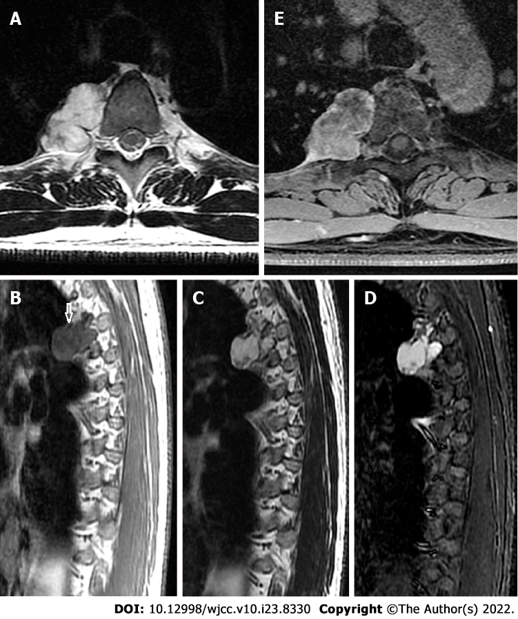
Figure 2 Magnetic resonance imaging.
A: T1-weighted magnetic resonance imaging in the transverse view showed a lobulated tumor on the right side of the thoracic vertebra; B and C: The lesion was bordered by a hypointense rim (arrow) and exhibited long T1 and T2 signals in the sagittal view on T1-weighted (B) and T2-weighted (C) imaging; D: Short tau inversion recovery imaging showed high signal intensity and mottling and patchy long T1 and short T2 abnormal signals within the mass; E: Enhancement was seen predominantly at the periphery of the lesion on post-enhanced images.
- Citation: Gao Y, Wang JG, Liu H, Gao CP. Periosteal chondroma of the rib: A case report. World J Clin Cases 2022; 10(23): 8330-8335
- URL: https://www.wjgnet.com/2307-8960/full/v10/i23/8330.htm
- DOI: https://dx.doi.org/10.12998/wjcc.v10.i23.8330


