Copyright
©The Author(s) 2022.
World J Clin Cases. Jul 26, 2022; 10(21): 7539-7544
Published online Jul 26, 2022. doi: 10.12998/wjcc.v10.i21.7539
Published online Jul 26, 2022. doi: 10.12998/wjcc.v10.i21.7539
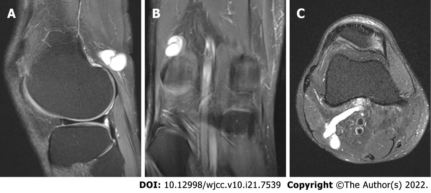
Figure 2 Magnetic resonance imaging views (sagittal, coronal, and axial) of the right knee revealing a hyperintense cystic mass with narrow and long stalk stretching out posterolaterally from middle popliteal fossa in supracondylar area.
A: Fat suppressed proton density sagittal magnetic resonance imaging (MRI) image; B: Fat suppressed proton density coronal MRI image; C: T2 pronton density axial MRI image.
- Citation: Won KH, Kang EY. Differential diagnosis and treatment of foot drop caused by an extraneural ganglion cyst above the knee: A case report. World J Clin Cases 2022; 10(21): 7539-7544
- URL: https://www.wjgnet.com/2307-8960/full/v10/i21/7539.htm
- DOI: https://dx.doi.org/10.12998/wjcc.v10.i21.7539


