Copyright
©The Author(s) 2022.
World J Clin Cases. Jul 26, 2022; 10(21): 7438-7444
Published online Jul 26, 2022. doi: 10.12998/wjcc.v10.i21.7438
Published online Jul 26, 2022. doi: 10.12998/wjcc.v10.i21.7438
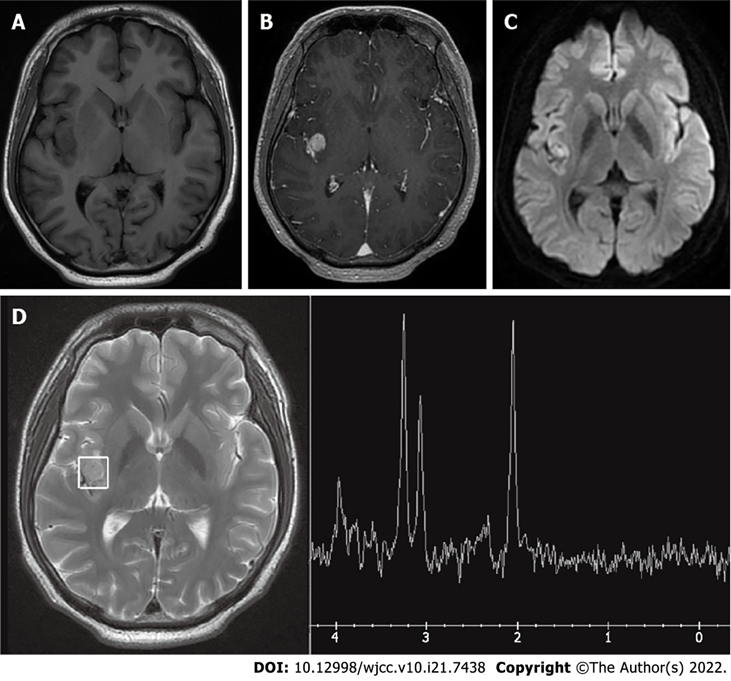
Figure 2 Magnetic resonance imaging and magnetic resonance spectroscopy before the first operation.
A and B: Axial T1-weighted magnetic resonance imaging scan demonstrating a hypointense right Sylvian fissure lesion, which was homogenously enhanced after contrast administration; C: Diffusion weighted imaging shows a hyperintense mass in the posterior part of the insular lobe; D: The magnetic resonance spectroscopy (MRS) N-acetyl aspartate (NAA) peak was slightly decreased.
- Citation: Wang A, Zhang X, Sun KK, Li C, Song ZM, Sun T, Wang F. Deep Sylvian fissure meningiomas: A case report. World J Clin Cases 2022; 10(21): 7438-7444
- URL: https://www.wjgnet.com/2307-8960/full/v10/i21/7438.htm
- DOI: https://dx.doi.org/10.12998/wjcc.v10.i21.7438


