Opinion Review Open Access
Copyright ©The Author(s) 2022. Published by Baishideng Publishing Group Inc. All rights reserved.
World J Clin Cases. Jul 26, 2022; 10(21): 7187-7194
Published online Jul 26, 2022. doi: 10.12998/wjcc.v10.i21.7187
Effects of glucocorticoids on leukocytes: Genomic and non-genomic mechanisms
Wan-Yu Jia, Jian-Jiang Zhang, Department of Pediatrics, The First Affiliated Hospital of Zhengzhou University, Zhengzhou 450052, Henan Province, China
Wan-Yu Jia, Jian-Jiang Zhang, Clinical Center of Pediatric Nephrology of Henan Province, The First Affiliated Hospital of Zhengzhou University, Zhengzhou 450052, Henan Province, China
ORCID number: Wan-Yu Jia (0000-0001-6543-0501); Jian-Jiang Zhang (0000-0003-2953-1924).
Author contributions: Zhang JJ conceived the idea for the manuscript; Jia WY reviewed the literature and drafted the manuscript; all authors have accepted responsibility for the entire content of this manuscript and approved its submission.
Supported by Provinces Co-construction Program of Medical Science and Technique Foundation of Henan Province, No. SB201901042; and Key Scientific Research Project of Colleges and Universities in Henan Province, No. 21A320070.
Conflict-of-interest statement: All the authors report no relevant conflicts of interest for this article.
Open-Access: This article is an open-access article that was selected by an in-house editor and fully peer-reviewed by external reviewers. It is distributed in accordance with the Creative Commons Attribution NonCommercial (CC BY-NC 4.0) license, which permits others to distribute, remix, adapt, build upon this work non-commercially, and license their derivative works on different terms, provided the original work is properly cited and the use is non-commercial. See: https://creativecommons.org/Licenses/by-nc/4.0/
Corresponding author: Jian-Jiang Zhang, Doctor, MD, PhD, Chief Physician, Department of Pediatrics, The First Affiliated Hospital of Zhengzhou University, No. 1 Jianshe East Road, Erqi District, Zhengzhou 450052, Henan Province, China. zhangjianjiang1@hotmail.com
Received: December 24, 2021
Peer-review started: December 24, 2021
First decision: February 14, 2022
Revised: February 21, 2022
Accepted: June 3, 2022
Article in press: June 3, 2022
Published online: July 26, 2022
Processing time: 199 Days and 5.9 Hours

Abstract

Glucocorticoids (GCs) have been widely used as immunosuppressants and anti-inflammatory agents to treat a variety of autoimmune and inflammatory diseases, and they fully exert their anti-inflammatory and immune-regulating effects in the body. The effect of GCs on white blood cells is an important part of their action. GCs can cause changes in peripheral blood white blood cell counts by regulating the proliferation, differentiation, and apoptosis of white blood cells. Although the total number of white blood cells, neutrophil counts, lymphocytes, and eosinophils increases, the counts of basic granulocytes and macrophages decreases. In addition, GCs can regulate the activation and secretion of white blood cells, inhibit the secretion of a variety of pro-inflammatory cytokines, the expression of chemokines, and promote the production of anti-inflammatory cytokines. For patients on GC therapy, the effects of GCs on leukocytes were similar to the changes in peripheral blood caused by bacterial infections. Thus, we suggest that clinicians should be more cautious in assessing the presence of infection in children with long-term use of GCs and avoid overuse of antibiotics in the presence of elevated leukocytes. GCs work through genomic and non-genomic mechanisms in the human body, which are mediated by GC receptors. In recent years, studies have not fully clarified the mechanism of GCs, and further research on these mechanisms will help to develop new therapeutic strategies.

Key Words: Glucocorticoid, Leukocyte, Count, Functions, Glucocorticoid receptor

Core Tip: Glucocorticoids (GCs) increase peripheral blood neutrophil counts through genomic and non-genomic actions and reduce the numbers of lymphocytes, eosinophils, basophils, and monocytes. GCs also regulate cell activity and affect cells. The functions of adhesion, activation, secretion and differentiation inhibit the expression of proinflammatory factors and chemokines. The mechanisms of action of GCs include effects on intracellular transcription and protein expression, effects on mitochondria, physical and chemical interactions with biological membranes, and receptor-mediated interactions with signal proteins.


INTRODUCTION

Glucocorticoids (GCs) are hormones produced by the adrenal cortex and can also be synthesized chemically. Exogenous hormones, including dexamethasone, prednisone, and methylprednisolone, are mainly used in diseases such as various allergic diseases and rheumatic immune diseases. Some GCs are derived from the adrenal cortex, such as hydrocortisone, which is mainly used for endocrine replacement therapy or as a treatment in various forms of congenital adrenal hyperplasia. GCs, which participate in many important physiological processes (such as metabolism, inflammation, immunity, and stress), are clinically used in the treatment of various types of acute and chronic inflammation, autoimmune diseases, organ transplantation, and tumors. GCs have many effects on immune regulation, and an important consideration is the influence on white blood cells, including the influence on their count and function. GCs act on nearly every cell type of the immune system, but the functional aspects of GCs differ by cell type. The mechanism of GC action on leukocytes is very complex, and studies have suggested that GCs exert anti-inflammatory and immunomodulatory effects through genomic and non-genomic mechanisms. Finally, 44 articles were selected for analysis and summary. This article summarizes the relevant research on the effects of GCs on leukocytes in recent decades.

EFFECTS OF GCs ON LEUKOCYTES: GENOMIC AND NON-GENOMIC MECHANISMS
Effects of GCs on neutrophils

Effects of GCs on neutrophil count: Barden et al[1] found that after 24 h of dexamethasone use in healthy volunteers, the neutrophil count increased in a dose-dependent manner[1]. GCs increase the neutrophil count in peripheral blood through a variety of mechanisms of action: (1) They promote neutrophil attachment to the blood vessel walls to enter the blood circulation (the marginal pool enters the circulating pool); (2) They reduce neutrophil outflow from the circulating pool; (3) They inhibit the apoptosis of neutrophils and delay their clearance in peripheral blood; and (4) They stimulate hematopoiesis in the bone marrow and increase the production of neutrophils in the peripheral blood[2,3].

Cavalcanti et al[4] confirmed in rat experiments that endogenous GCs can promote the maturation of bone marrow neutrophils and the mobilization of neutrophils from the bone marrow into the circulation[4]. A few studies have confirmed that GCs can inhibit the apoptosis of neutrophils and delay their clearance both in vivo and in vitro[3,5]. However, the mechanism by which GCs regulate neutrophil apoptosis is still unclear. It has been suggested that GC-mediated apoptosis inhibition mechanisms may include upregulation of antiapoptotic B-cell lymphoma-2 (Bcl-2) and members of the inhibitor of apoptosis family. For example, dexamethasone has been shown to induce survival and enhance McL-1, a proapoptotic member of the Bcl-2 family, through phosphatidylinositol 3-kinase (PI3K) and P38 mitogen-activated protein kinase (P38 MAPK) in human neutrophils[6]. Research by Chapman et al[7] confirmed that GCs can promote macrophages to phagocytose apoptotic leukocytes and cause them to rapidly degrade without causing proinflammatory secretory reactions. This effect can be strengthened by 11β-hydroxysteroid dehydrogenase[7]. Additionally, GCs can stimulate bone marrow hematopoiesis and increase bone marrow neutrophil production. Endogenous GCs are one of the factors stimulating the maturation of bone marrow neutrophils and promoting the mobilization of neutrophils from the bone marrow into the circulation[3]. Human and mouse neutrophil migration depends on the induction of interleukin (IL)-8 expression, and this induction is inhibited by GCs[8]. For the migration of neutrophils, GCs inhibit this migration by attenuating the expression of CXC receptor 2 agonists such as IL-8 and CXCL18b[9]. Ricci et al[10] found in mouse experiments that GC-induced leucine zipper (GILZ) inhibited the migration of neutrophils by controlling the expression of annexin A1[10].

Effects of GCs on the intercellular adhesion of neutrophils: GCs can inhibit neutrophil-endothelial cell adhesion, and their molecular mechanism may include downregulation of GCs on cell surface adhesion factors, such as intercellular adhesion molecule-1 (ICAM-1), endothelial cell adhesion molecule-1, E-selectin, P-selectin and L-selectin, thereby affecting the leukocyte-endothelial cell interaction[4]. The stagnation of leukocytes on the endothelial surface is largely mediated by leukocyte integrins, especially β1 (late antigen-4) and β2 (lymphatic function-related antigen-1 and macrophage antigen-1), and their respective endothelial counterparts are cell adhesion molecule-1 (CAM-1), ICAM-1 and ICAM-2. In the process of inflammation, CAM, ICAM-1, and E-selectin, among other adhesion molecules, are significantly upregulated to promote the adhesion, aggregation and activation of leukocytes. Cell experiments have confirmed that GCs can inhibit the upregulation of these factors, thereby inhibiting cell adhesion. Therefore, GCs may also prohibit the expression of adhesion molecules by inhibiting the synthesis of cytokines. Most of the effects of GCs are caused by genomic mechanisms; that is, they affect cell transcription and protein expression, such as inhibiting the activation of the nuclear factor-κB (NF-κB) pathway and inducing MAPK phosphatase-1 to inhibit MAPK activation, thereby reducing the expression of cytokines, chemokines and adhesion molecules[11-13], including CD44 and integrin lymphocyte function-associated antigen 1 (LFA-1) and very late antigen 4, to inhibit neutrophil adhesion[14].

Effects of GCs on neutrophil function: Neutrophils are an important line of defense in the human body against foreign pathogens. Neutrophils are rapidly activated after encountering foreign antigens (such as viruses or bacteria), and their activation is followed by phagocytosis and degranulation. Then, enzymes in the particles enter the phagolysosome or cytoplasm. These enzymes are excreted outside of the cell and exert functions such as sterilization, lysis, and digestion of foreign bodies. In vitro studies have found that when human neutrophils are acutely exposed to methylprednisolone or hydrocortisone, their N-formyl-methionyl-leucyl-phenylalanine-induced neutrophil degranulation is obviously suppressed, and this effect is not influenced by RU486 or cycloheximide[15]. Research by Ricci et al[16] confirmed that GILZ inhibits the activation and migration of neutrophils to inflammation sites by inhibiting the MAPK pathway and the secretion of proinflammatory cytokines by neutrophils[16].

Effects of GCs on lymphocytes

Effects of GCs on lymphocyte count: GCs can reduce peripheral blood lymphocyte counts. The peripheral blood lymphocyte count decreased significantly after short- or long-term application of GCs in both animal and clinical experiments[17]. GCs promote the apoptosis of lymphocytes and significantly decrease the lymphocyte count in peripheral blood. In vitro cell culture of mouse spleen cells and bone marrow lymphocytes revealed that dexamethasone stimulated the apoptosis of all B cell developmental subgroups, while in vivo experiments showed that immature B cells promoted GCs. Multiple injections of dexamethasone regulated the number of B cells in the bone marrow but did not affect the number of mature B cells in the body[18]. Costa et al[19] also confirmed that hydrocortisone could regulate the production of B lymphocytes[19]. Several experiments have shown that GCs are involved in regulating T cell apoptosis, and the mechanisms include genomic and nongenomic mechanisms. GCs exert their effects predominantly through the GC glucocorticoid receptor (GR). The genomic function of GCs is mainly to bind to specific GRs in the cytoplasm to form complexes and transfer to the nucleus, thus regulating the transcriptional activity of GC response genes. The GR can dimerize and directly bind DNA at GC response elements, affecting transcription rates. In addition, ligand-bound GR can be recruited to specific genomic sites via protein-protein interactions with other DNA-bound transcription factors. GCs also exert genomic effects by interfering with the activity of transcription factors and signaling molecules[20]. Genes with up- or down-regulated expression in GC-induced apoptosis include c-myc, tdag8, dig2, Bim, and PUMA[21,22]. Non-genomic effects include the physicochemical interactions of GCs with biological membranes, the effects mediated by the GC-GR complex and the GC-induced mitochondrial apoptotic pathway. These mechanisms have not been fully elucidated[21]. Multiple mouse experiments have demonstrated that pro- and antiapoptotic members of the Bcl-2 family are involved in GC-induced apoptosis in lymphocytes. Caspase-3 and caspase-8 are thought to mediate GC-induced apoptosis[21-24]. The events involved in GC-induced apoptosis include the production of ceramide, changes in intracellular sodium and potassium levels, the activation of PI3K and inositol triphosphate receptors, and the interaction of GR and other signaling proteins, such as protein kinase C and Raf[22].

Effects of GCs on lymphocyte activity: GCs can inhibit lymphocyte proliferation and reduce lymphocyte activity, thereby inhibiting cellular and humoral immunity. GCs affect the activity of transcription factors downstream of T cell receptor (TCR) activation, including NF-κB, activator protein 1 (AP-1) and nuclear factor of activated T cells[14,22]. GCs can also regulate T cell activation by regulating the functions of DCs, macrophages, and mast cells[25]. Studies have suggested that the effect of GCs on T cells is partly mediated by GILZ. GILZ regulates cell apoptosis, proliferation and differentiation by regulating transcription factors and signaling pathways related to host immunity and inflammation. For example, GILZ associates with NF-κB and inhibits NF-κB- and AP-1-dependent transcription. GILZ also binds Raf and Ras and inhibits the activation of Ras/Raf downstream targets, including MAPK1. GILZ also promotes the activity of regulatory T cells (Tregs) by activating transforming growth factor-β signaling. Ultimately, these effects inhibit T cell activation, regulate T helper Th-1, Th-2, and Th-17 cell differentiation, and reduce interferon-γ (IFN-γ) production by Th1, CD8 T, and NK cells, leading to the inhibition of cytotoxic responses[25,26]. A large number of studies have shown that GCs preferentially inhibit the responses of Th-1 cells and Th-17 cells while retaining or even promoting the functions of Th-2 cells and regulatory T cells[14]. GCs have the potential to promote Th2 cytokine production. CD4 T cells pretreated with dexamethasone produce higher levels of IL-4, IL-10 and IL-13[26]. GCs can inhibit the adhesion of lymphocytes to endothelial cell lines and inhibit the intercellular aggregation of activated lymphocytes[11]. Xing et al[27] proposed that GCs induce programmed cell death 1 (PD-1) expression in activated T cells and inhibit TCR-mediated T cell proliferation and cytokine production, including IL-2, IFN-γ and tumour necrosis factor-α (TNF-α)[27]. Studies by Okoye et al[28] confirmed that dexamethasone can affect the activity of T cells by promoting the expression of PD-1 and CTLA-4 through activated T cells, inhibiting the secretion of cytokines and inducing their apoptosis[28]. GCs can regulate the maturation and differentiation of regulatory T cell subsets. For patients with autoimmune diseases, allergies or autoinflammatory diseases, GC therapy can lead to the expansion of Treg cells[29]. Cain et al[30] found through experiments in mice that GCs regulate the expression of CXCR4 in B cells, thereby promoting their migration to the bone marrow[30].

Effects of GCs on macrophages

Effects of GCs on macrophage count: GCs have a concentration-dependent dual effect on macrophages. Low concentrations have immunostimulatory effects on macrophage functions such as adhesion, transformation, phagocytosis and cytokine production, while high concentrations exert immunosuppressive effects[31,32]. GCs can also directly induce specific changes in cell survival, proliferation and phagocytosis, thereby inhibiting cell proliferation. Ai et al[5] found that dexamethasone induced GR recruitment to the transcription factor Krüppel-like factor 9 promoter and increased mitochondrial ROS production, leading to mitochondrial-dependent apoptosis of macrophages[5].

Effects of GCs on macrophage differentiation: GCs affect the typing and differentiation of macrophages. Heideveld et al[33] confirmed that GC receptor activation differentiates monocytes into anti-inflammatory tissue macrophages with an M2 phenotype[33]. Experiments have confirmed that GCs can stimulate human and mouse macrophages to phagocytose apoptotic substances, and monocytes can change their intracellular composition under the induction of GCs, regulate cell skeletal reorganization and adhesion, and thus transform into a highly phagocytic monocyte-derived macrophage (MDMφ) phenotype[31]. In animal models of arthritis and acute lung injury, GCs have been shown to inhibit the differentiation of macrophages to the M1 phenotype[9].

Effects of GCs on macrophage function: GCs can stimulate the ability of macrophages to swallow apoptotic substances. Exposure of mouse and human macrophages to GCs for 24 h leads to increased uptake of apoptotic bodies[34]. GCs initiate gene programs in monocytes and macrophages to promote the phagocytosis of apoptotic cells and debridement cells[14]. GCs have been shown to inhibit the production of several proinflammatory cytokines in human monocytes and macrophages, including IL-1β, IL-6, IL-12, TNF-α, and granulocyte-macrophage colony-stimulating factor (GM-CSF)[35].

Effects of GCs on eosinophils

Effects of GCs on eosinophil count: GC application can reduce the number of peripheral blood eosinophils. The application of inhaled corticosteroids can reduce the number of eosinophils in the peripheral blood circulation and tracheal mucosa of asthma patients[36]. When GCs are used in Crohn's disease, chronic obstructive pulmonary disease, eosinophilic bronchitis, eosinophilic gastroenteritis, nephrotic syndrome and other diseases, a decrease in the patient's eosinophil count can be observed[37-39]. GCs can promote the apoptosis of eosinophils[9,31]. Cell experiments indicate that without the involvement of cytokines, GCs can accelerate the apoptosis of eosinophils, while GCs can reverse the cell survival induced by TNF-α and antagonize low-dose GM-CSF and IL-5 inhibition of apoptosis on eosinophils but cannot reverse the effects induced by IL-3, IL-5, and GM-CSF at the concentration that produces the maximum anti-apoptotic effect[40]. The findings of Hong et al[41] showed that GC-induced eosinophilia is caused by CXCR4-dependent migration of eosinophils to the bone marrow[41].

Effects of GCs on eosinophil function: In mouse eosinophils, dexamethasone and budesonide reduce the expression of basal CD11b in a concentration-dependent manner, thereby affecting the adhesion of eosinophils[42]. A study found that preincubation of cells with different concentrations of budesonide can also effectively downregulate the expression of LFA-1 and Mac-1 induced by GM-CSF on eosinophils and downregulate the migration of eosinophils through airway epithelial cells[43]. Studies have found that the level of IL-5 in sputum decreases after prednisone or prednisolone treatment, further affecting the recruitment, activation and survival of eosinophils[37].

Effects of GCs on basophils

GCs can cause a decrease in peripheral blood basophil count. Barden et al[1] found that peripheral blood basophil hormone decreased significantly after 4 h of dexamethasone use in healthy volunteers[1]. GCs can inhibit the release of histamine from basophils, increase the transcription of leukocyte protease inhibitors, and reduce the basophil count[31]. Thus, GCs can promote the apoptosis of basophils[9].

Effects of GCs on dendritic cells

Both in vivo and in vitro, GCs can inhibit dendritic cell (DC) maturation and weaken the activity of DCs[31]. Studies have shown that the application of inhaled corticosteroids can rapidly reduce the number of peripheral blood DCs in patients with allergic rhinitis, and the DC activation markers CD86 and CD80 are reduced to varying degrees, suggesting that GCs inhibit DC activation[44]. GCs inhibit DC function, reduce the expression of class II MHC and costimulatory molecules, reduce proinflammatory cytokines and increase the secretion of anti-inflammatory cytokines. GCs can also improve the ability of DCs to capture antigens but inhibit their function as antigen-presenting cells[25,45].

The role of GCs on leukocytes is very important, and a lack of GCs or excessive GCs in the body will cause abnormal states of the body. Impaired adrenocortical axis integrity in vivo can lead to immunodeficiencies. For example, patients with deficits in anterior pituitary function and variable immune deficiency presenting with adrenocorticotropin deficiency have decreased B cells, persistent hypoglobulinemia, and susceptibility to infection. The patient's symptoms improved significantly after hydrocortisone replacement therapy[46,47]. Elevated leukocytes and neutrophils and decreased lymphocytes can be observed in patients with Cushing's syndrome. During remission or hormonal control of the disease, a significant decrease in neutrophil counts and in the hemoglobin concentration together with a rise in lymphocyte numbers was observed[48].

CONCLUSION

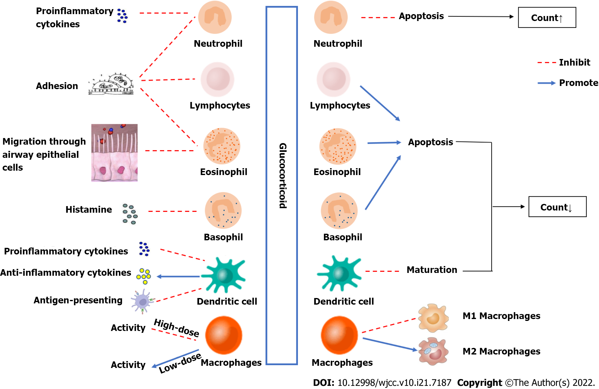
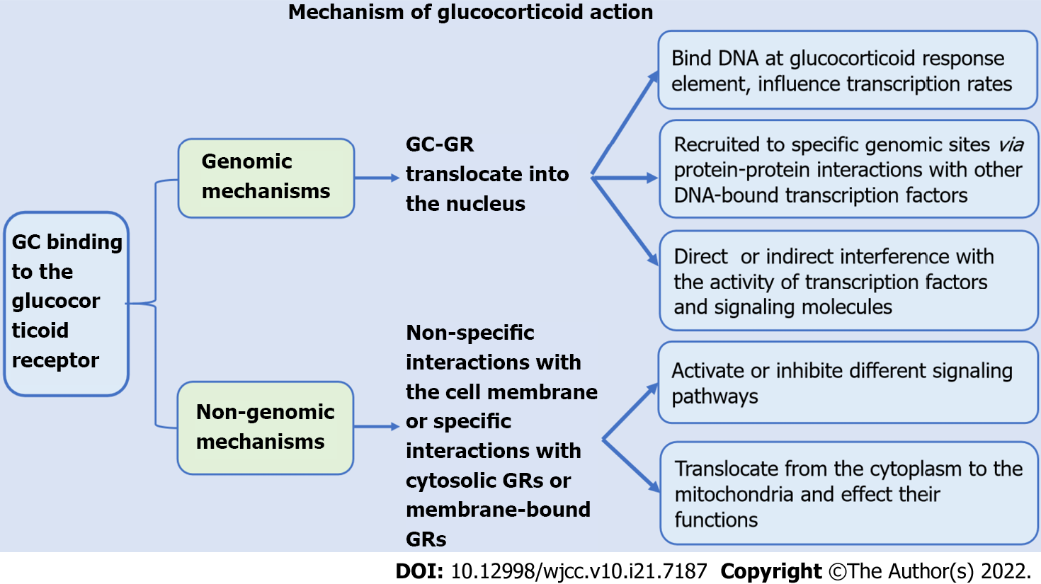
GCs increase peripheral blood neutrophil counts through genomic and non-genomic mechanisms and inhibit cell adhesion and neutrophil activation and secretion. Moreover, GCs reduce the counts of lymphocytes, eosinophils, basophils, and mononuclear macrophages, reduce cell activity, regulate the distribution of T cell subsets, and inhibit the expression of proinflammatory factors and chemokines. GCs increase peripheral blood neutrophil counts through genomic and non-genomic effects and reduce the numbers of lymphocytes, eosinophils, basophils, and monocytes. GCs also regulate cell activity and affect cells. The processes of adhesion, activation, secretion and differentiation inhibit the expression of proinflammatory factors and chemokines (Figure 1). The mechanisms of action of GCs include effects on intracellular transcription and protein expression, effects on mitochondria, physical and chemical interactions with biological membranes, and receptor-mediated interactions with signal proteins (Figure 2). Clinically, due to the immunosuppressive effect of GCs, patients treated with GCs are prone to coinfection. For patients on GC therapy, the effects of GCs on white blood cells are similar to the effects of bacterial infections on white blood cells, which may lead to misdiagnosis of patients suffering from infection and overuse of antibiotics. Therefore, it is very important to identify whether patients who use GCs have infections. As a consequence, we suggest that clinicians should be more cautious in assessing the presence of infection in children with long-term use of GCs and avoid overuse of antibiotics in the presence of elevated leukocytes. The role of GCs is very important, and a lack of or excess of GCs in the body can cause abnormalities. In recent years, many studies have examined intracellular transcription and the cellular pathways related to the effects of GCs on white blood cells, but these processes are not yet fully clear. Further research on these mechanisms will help to develop new therapeutic strategies.

Figure 1
Figure 1 Mechanism of glucocorticoid action.
Figure 2
Figure 2 Effects of glucocorticoids on leukocytes. GC: Glucocorticoid; GR: Glucocorticoid receptor.
Footnotes

Provenance and peer review: Unsolicited article; Externally peer reviewed.

Peer-review model: Single blind

Specialty type: Medicine, research and experimental

Country/Territory of origin: China

Peer-review report’s scientific quality classification

Grade A (Excellent): 0

Grade B (Very good): B

Grade C (Good): C, C

Grade D (Fair): D, D, D

Grade E (Poor): 0

P-Reviewer: Amornyotin S, Thailand; Aydin S, Turkey; Dauyey K, Kazakhstan; Moshref RH, Saudi Arabia; Novita BD, Indonesia A-Editor: Liu X, China S-Editor: Fan JR L-Editor: Webster JR P-Editor: Fan JR

References
1.  Barden A, Phillips M, Hill LM, Fletcher EM, Mas E, Loh PS, French MA, Ho KM, Mori TA, Corcoran TB. Antiemetic doses of dexamethasone and their effects on immune cell populations and plasma mediators of inflammation resolution in healthy volunteers. Prostaglandins Leukot Essent Fatty Acids. 2018;139:31-39.  [PubMed]  [DOI]  [Cited in This Article: ]  [Cited by in Crossref: 11]  [Cited by in F6Publishing: 17]  [Article Influence: 2.8]  [Reference Citation Analysis (0)]
2.  Velthove KJ, Leufkens HG, Souverein PC, Schweizer RC, Bracke M, van Solinge WW. Effects of glucocorticoids on the neutrophil count: a cohort study among hospitalized patients. Pulm Pharmacol Ther. 2010;23:129-134.  [PubMed]  [DOI]  [Cited in This Article: ]  [Cited by in Crossref: 9]  [Cited by in F6Publishing: 9]  [Article Influence: 0.6]  [Reference Citation Analysis (1)]
3.  Ronchetti S, Ricci E, Migliorati G, Gentili M, Riccardi C. How Glucocorticoids Affect the Neutrophil Life. Int J Mol Sci. 2018;19.  [PubMed]  [DOI]  [Cited in This Article: ]  [Cited by in Crossref: 85]  [Cited by in F6Publishing: 117]  [Article Influence: 19.5]  [Reference Citation Analysis (2)]
4.  Cavalcanti DM, Lotufo CM, Borelli P, Ferreira ZS, Markus RP, Farsky SH. Endogenous glucocorticoids control neutrophil mobilization from bone marrow to blood and tissues in non-inflammatory conditions. Br J Pharmacol. 2007;152:1291-1300.  [PubMed]  [DOI]  [Cited in This Article: ]  [Cited by in Crossref: 53]  [Cited by in F6Publishing: 42]  [Article Influence: 2.5]  [Reference Citation Analysis (0)]
5.  Ai F, Zhao G, Lv W, Liu B, Lin J. Dexamethasone induces aberrant macrophage immune function and apoptosis. Oncol Rep. 2020;43:427-436.  [PubMed]  [DOI]  [Cited in This Article: ]  [Cited by in Crossref: 13]  [Cited by in F6Publishing: 21]  [Article Influence: 4.2]  [Reference Citation Analysis (0)]
6.  Hirsch G, Lavoie-Lamoureux A, Beauchamp G, Lavoie JP. Neutrophils are not less sensitive than other blood leukocytes to the genomic effects of glucocorticoids. PLoS One. 2012;7:e44606.  [PubMed]  [DOI]  [Cited in This Article: ]  [Cited by in Crossref: 40]  [Cited by in F6Publishing: 42]  [Article Influence: 3.5]  [Reference Citation Analysis (0)]
7.  Chapman KE, Coutinho A, Gray M, Gilmour JS, Savill JS, Seckl JR. Local amplification of glucocorticoids by 11beta-hydroxysteroid dehydrogenase type 1 and its role in the inflammatory response. Ann N Y Acad Sci. 2006;1088:265-273.  [PubMed]  [DOI]  [Cited in This Article: ]  [Cited by in Crossref: 52]  [Cited by in F6Publishing: 52]  [Article Influence: 3.1]  [Reference Citation Analysis (0)]
8.  Huang G, Liang B, Liu G, Liu K, Ding Z. Low dose of glucocorticoid decreases the incidence of complications in severely burned patients by attenuating systemic inflammation. J Crit Care. 2015;30:436.e7-436.11.  [PubMed]  [DOI]  [Cited in This Article: ]  [Cited by in Crossref: 10]  [Cited by in F6Publishing: 9]  [Article Influence: 0.9]  [Reference Citation Analysis (0)]
9.  Xie Y, Tolmeijer S, Oskam JM, Tonkens T, Meijer AH, Schaaf MJM. Glucocorticoids inhibit macrophage differentiation towards a pro-inflammatory phenotype upon wounding without affecting their migration. Dis Model Mech. 2019;12.  [PubMed]  [DOI]  [Cited in This Article: ]  [Cited by in Crossref: 46]  [Cited by in F6Publishing: 47]  [Article Influence: 9.4]  [Reference Citation Analysis (0)]
10.  Ricci E, Ronchetti S, Pericolini E, Gabrielli E, Cari L, Gentili M, Roselletti E, Migliorati G, Vecchiarelli A, Riccardi C. Role of the glucocorticoid-induced leucine zipper gene in dexamethasone-induced inhibition of mouse neutrophil migration via control of annexin A1 expression. FASEB J. 2017;31:3054-3065.  [PubMed]  [DOI]  [Cited in This Article: ]  [Cited by in Crossref: 29]  [Cited by in F6Publishing: 30]  [Article Influence: 4.3]  [Reference Citation Analysis (0)]
11.  Pitzalis C, Pipitone N, Perretti M. Regulation of leukocyte-endothelial interactions by glucocorticoids. Ann N Y Acad Sci. 2002;966:108-118.  [PubMed]  [DOI]  [Cited in This Article: ]  [Cited by in Crossref: 106]  [Cited by in F6Publishing: 109]  [Article Influence: 5.0]  [Reference Citation Analysis (0)]
12.  Gregory JL, Hall P, Leech M, Morand EF, Hickey MJ. Independent roles of macrophage migration inhibitory factor and endogenous, but not exogenous glucocorticoids in regulating leukocyte trafficking. Microcirculation. 2009;16:735-748.  [PubMed]  [DOI]  [Cited in This Article: ]  [Cited by in Crossref: 18]  [Cited by in F6Publishing: 20]  [Article Influence: 1.4]  [Reference Citation Analysis (0)]
13.  Nakagawa M, Bondy GP, Waisman D, Minshall D, Hogg JC, van Eeden SF. The effect of glucocorticoids on the expression of L-selectin on polymorphonuclear leukocyte. Blood. 1999;93:2730-2737.  [PubMed]  [DOI]  [Cited in This Article: ]
14.  Cain DW, Cidlowski JA. Immune regulation by glucocorticoids. Nat Rev Immunol. 2017;17:233-247.  [PubMed]  [DOI]  [Cited in This Article: ]  [Cited by in Crossref: 737]  [Cited by in F6Publishing: 967]  [Article Influence: 138.1]  [Reference Citation Analysis (0)]
15.  Panettieri RA, Schaafsma D, Amrani Y, Koziol-White C, Ostrom R, Tliba O. Non-genomic Effects of Glucocorticoids: An Updated View. Trends Pharmacol Sci. 2019;40:38-49.  [PubMed]  [DOI]  [Cited in This Article: ]  [Cited by in Crossref: 96]  [Cited by in F6Publishing: 143]  [Article Influence: 23.8]  [Reference Citation Analysis (0)]
16.  Ricci E, Ronchetti S, Gabrielli E, Pericolini E, Gentili M, Roselletti E, Vecchiarelli A, Riccardi C. GILZ restrains neutrophil activation by inhibiting the MAPK pathway. J Leukoc Biol. 2019;105:187-194.  [PubMed]  [DOI]  [Cited in This Article: ]  [Cited by in Crossref: 27]  [Cited by in F6Publishing: 27]  [Article Influence: 4.5]  [Reference Citation Analysis (0)]
17.  Ohkaru Y, Arai N, Ohno H, Sato S, Sakakibara Y, Suzuki H, Aritoshi S, Akimoto S, Ban T, Tanihata J, Tachiyashiki K, Imaizumi K. Acute and Subacute Effects of Dexamethasone on the Number of White Blood Cells in Rats. J Health Sci. 2010;56:215-220.  [PubMed]  [DOI]  [Cited in This Article: ]
18.  Gruver-Yates AL, Quinn MA, Cidlowski JA. Analysis of glucocorticoid receptors and their apoptotic response to dexamethasone in male murine B cells during development. Endocrinology. 2014;155:463-474.  [PubMed]  [DOI]  [Cited in This Article: ]  [Cited by in Crossref: 57]  [Cited by in F6Publishing: 54]  [Article Influence: 5.4]  [Reference Citation Analysis (0)]
19.  Costa KMD, Valente RC, Silva JMCD, Paiva LS, Rumjanek VM. Glucocorticoid susceptibility and in vivo ABCB1 activity differ in murine B cell subsets. An Acad Bras Cienc. 2018;90:3081-3097.  [PubMed]  [DOI]  [Cited in This Article: ]  [Cited by in Crossref: 3]  [Cited by in F6Publishing: 4]  [Article Influence: 1.0]  [Reference Citation Analysis (0)]
20.  Franco LM, Gadkari M, Howe KN, Sun J, Kardava L, Kumar P, Kumari S, Hu Z, Fraser IDC, Moir S, Tsang JS, Germain RN. Immune regulation by glucocorticoids can be linked to cell type-dependent transcriptional responses. J Exp Med. 2019;216:384-406.  [PubMed]  [DOI]  [Cited in This Article: ]  [Cited by in Crossref: 88]  [Cited by in F6Publishing: 112]  [Article Influence: 22.4]  [Reference Citation Analysis (0)]
21.  Prenek L, Boldizsár F, Kugyelka R, Ugor E, Berta G, Németh P, Berki T. The regulation of the mitochondrial apoptotic pathway by glucocorticoid receptor in collaboration with Bcl-2 family proteins in developing T cells. Apoptosis. 2017;22:239-253.  [PubMed]  [DOI]  [Cited in This Article: ]  [Cited by in Crossref: 38]  [Cited by in F6Publishing: 36]  [Article Influence: 6.0]  [Reference Citation Analysis (0)]
22.  Herold MJ, McPherson KG, Reichardt HM. Glucocorticoids in T cell apoptosis and function. Cell Mol Life Sci. 2006;63:60-72.  [PubMed]  [DOI]  [Cited in This Article: ]  [Cited by in Crossref: 282]  [Cited by in F6Publishing: 227]  [Article Influence: 12.6]  [Reference Citation Analysis (0)]
23.  Harr MW, Caimi PF, McColl KS, Zhong F, Patel SN, Barr PM, Distelhorst CW. Inhibition of Lck enhances glucocorticoid sensitivity and apoptosis in lymphoid cell lines and in chronic lymphocytic leukemia. Cell Death Differ. 2010;17:1381-1391.  [PubMed]  [DOI]  [Cited in This Article: ]  [Cited by in Crossref: 47]  [Cited by in F6Publishing: 48]  [Article Influence: 3.4]  [Reference Citation Analysis (0)]
24.  Kiuchi Z, Nishibori Y, Kutsuna S, Kotani M, Hada I, Kimura T, Fukutomi T, Fukuhara D, Ito-Nitta N, Kudo A, Takata T, Ishigaki Y, Tomosugi N, Tanaka H, Matsushima S, Ogasawara S, Hirayama Y, Takematsu H, Yan K. GLCCI1 is a novel protector against glucocorticoid-induced apoptosis in T cells. FASEB J. 2019;33:7387-7402.  [PubMed]  [DOI]  [Cited in This Article: ]  [Cited by in Crossref: 9]  [Cited by in F6Publishing: 9]  [Article Influence: 1.8]  [Reference Citation Analysis (0)]
25.  Cannarile L, Delfino DV, Adorisio S, Riccardi C, Ayroldi E. Implicating the Role of GILZ in Glucocorticoid Modulation of T-Cell Activation. Front Immunol. 2019;10:1823.  [PubMed]  [DOI]  [Cited in This Article: ]  [Cited by in Crossref: 29]  [Cited by in F6Publishing: 32]  [Article Influence: 6.4]  [Reference Citation Analysis (0)]
26.  Shimba A, Ikuta K. Glucocorticoids Regulate Circadian Rhythm of Innate and Adaptive Immunity. Front Immunol. 2020;11:2143.  [PubMed]  [DOI]  [Cited in This Article: ]  [Cited by in Crossref: 28]  [Cited by in F6Publishing: 28]  [Article Influence: 7.0]  [Reference Citation Analysis (0)]
27.  Xing K, Gu B, Zhang P, Wu X. Dexamethasone enhances programmed cell death 1 (PD-1) expression during T cell activation: an insight into the optimum application of glucocorticoids in anti-cancer therapy. BMC Immunol. 2015;16:39.  [PubMed]  [DOI]  [Cited in This Article: ]  [Cited by in Crossref: 74]  [Cited by in F6Publishing: 91]  [Article Influence: 10.1]  [Reference Citation Analysis (0)]
28.  Okoye IS, Xu L, Walker J, Elahi S. The glucocorticoids prednisone and dexamethasone differentially modulate T cell function in response to anti-PD-1 and anti-CTLA-4 immune checkpoint blockade. Cancer Immunol Immunother. 2020;69:1423-1436.  [PubMed]  [DOI]  [Cited in This Article: ]  [Cited by in Crossref: 22]  [Cited by in F6Publishing: 33]  [Article Influence: 8.3]  [Reference Citation Analysis (0)]
29.  Cari L, De Rosa F, Nocentini G, Riccardi C. Context-Dependent Effect of Glucocorticoids on the Proliferation, Differentiation, and Apoptosis of Regulatory T Cells: A Review of the Empirical Evidence and Clinical Applications. Int J Mol Sci. 2019;20.  [PubMed]  [DOI]  [Cited in This Article: ]  [Cited by in Crossref: 38]  [Cited by in F6Publishing: 35]  [Article Influence: 7.0]  [Reference Citation Analysis (0)]
30.  Cain DW, Bortner CD, Diaz-Jimenez D, Petrillo MG, Gruver-Yates A, Cidlowski JA. Murine Glucocorticoid Receptors Orchestrate B Cell Migration Selectively between Bone Marrow and Blood. J Immunol. 2020;205:619-629.  [PubMed]  [DOI]  [Cited in This Article: ]  [Cited by in Crossref: 12]  [Cited by in F6Publishing: 19]  [Article Influence: 4.8]  [Reference Citation Analysis (0)]
31.  Zen M, Canova M, Campana C, Bettio S, Nalotto L, Rampudda M, Ramonda R, Iaccarino L, Doria A. The kaleidoscope of glucorticoid effects on immune system. Autoimmun Rev. 2011;10:305-310.  [PubMed]  [DOI]  [Cited in This Article: ]  [Cited by in Crossref: 171]  [Cited by in F6Publishing: 194]  [Article Influence: 14.9]  [Reference Citation Analysis (0)]
32.  Lim HY, Müller N, Herold MJ, van den Brandt J, Reichardt HM. Glucocorticoids exert opposing effects on macrophage function dependent on their concentration. Immunology. 2007;122:47-53.  [PubMed]  [DOI]  [Cited in This Article: ]  [Cited by in Crossref: 140]  [Cited by in F6Publishing: 141]  [Article Influence: 8.3]  [Reference Citation Analysis (0)]
33.  Heideveld E, Hampton-O'Neil LA, Cross SJ, van Alphen FPJ, van den Biggelaar M, Toye AM, van den Akker E. Glucocorticoids induce differentiation of monocytes towards macrophages that share functional and phenotypical aspects with erythroblastic island macrophages. Haematologica. 2018;103:395-405.  [PubMed]  [DOI]  [Cited in This Article: ]  [Cited by in Crossref: 44]  [Cited by in F6Publishing: 46]  [Article Influence: 6.6]  [Reference Citation Analysis (0)]
34.  Giles KM, Ross K, Rossi AG, Hotchin NA, Haslett C, Dransfield I. Glucocorticoid augmentation of macrophage capacity for phagocytosis of apoptotic cells is associated with reduced p130Cas expression, loss of paxillin/pyk2 phosphorylation, and high levels of active Rac. J Immunol. 2001;167:976-986.  [PubMed]  [DOI]  [Cited in This Article: ]  [Cited by in Crossref: 125]  [Cited by in F6Publishing: 129]  [Article Influence: 5.6]  [Reference Citation Analysis (0)]
35.  Ehrchen JM, Roth J, Barczyk-Kahlert K. More Than Suppression: Glucocorticoid Action on Monocytes and Macrophages. Front Immunol. 2019;10:2028.  [PubMed]  [DOI]  [Cited in This Article: ]  [Cited by in Crossref: 69]  [Cited by in F6Publishing: 107]  [Article Influence: 21.4]  [Reference Citation Analysis (0)]
36.  Lommatzsch M, Klein M, Stoll P, Virchow JC. Impact of an increase in the inhaled corticosteroid dose on blood eosinophils in asthma. Thorax. 2019;74:417-418.  [PubMed]  [DOI]  [Cited in This Article: ]  [Cited by in Crossref: 21]  [Cited by in F6Publishing: 31]  [Article Influence: 5.2]  [Reference Citation Analysis (0)]
37.  Sakae TM, Maurici R, Trevisol DJ, Pizzichini MM, Pizzichini E. Effects of prednisone on eosinophilic bronchitis in asthma: a systematic review and meta-analysis. J Bras Pneumol. 2014;40:552-563.  [PubMed]  [DOI]  [Cited in This Article: ]  [Cited by in Crossref: 5]  [Cited by in F6Publishing: 5]  [Article Influence: 0.6]  [Reference Citation Analysis (0)]
38.  Higham A, Scott T, Li J, Gaskell R, Dikwa AB, Shah R, Montero-Fernandez MA, Lea S, Singh D. Effects of corticosteroids on COPD lung macrophage phenotype and function. Clin Sci (Lond). 2020;134:751-763.  [PubMed]  [DOI]  [Cited in This Article: ]  [Cited by in Crossref: 14]  [Cited by in F6Publishing: 19]  [Article Influence: 4.8]  [Reference Citation Analysis (0)]
39.  Xue J, Cui YN, Chen P, Cai S, Chen L, Dai ZS, Chen Y. [Blood eosinophils: a biomarker of response to glucocorticoids and increased readmissions in severe hospitalized exacerbations of COPD]. Zhonghua Jie He He Hu Xi Za Zhi. 2019;42:426-431.  [PubMed]  [DOI]  [Cited in This Article: ]  [Cited by in F6Publishing: 4]  [Reference Citation Analysis (1)]
40.  Meagher LC, Cousin JM, Seckl JR, Haslett C. Opposing effects of glucocorticoids on the rate of apoptosis in neutrophilic and eosinophilic granulocytes. J Immunol. 1996;156:4422-4428.  [PubMed]  [DOI]  [Cited in This Article: ]
41.  Hong SG, Sato N, Legrand F, Gadkari M, Makiya M, Stokes K, Howe KN, Yu SJ, Linde NS, Clevenger RR, Hunt T, Hu Z, Choyke PL, Dunbar CE, Klion AD, Franco LM. Glucocorticoid-induced eosinopenia results from CXCR4-dependent bone marrow migration. Blood. 2020;136:2667-2678.  [PubMed]  [DOI]  [Cited in This Article: ]  [Cited by in Crossref: 10]  [Cited by in F6Publishing: 17]  [Article Influence: 5.7]  [Reference Citation Analysis (0)]
42.  Lim LH, Flower RJ, Perretti M, Das AM. Glucocorticoid receptor activation reduces CD11b and CD49d levels on murine eosinophils: characterization and functional relevance. Am J Respir Cell Mol Biol. 2000;22:693-701.  [PubMed]  [DOI]  [Cited in This Article: ]  [Cited by in Crossref: 25]  [Cited by in F6Publishing: 26]  [Article Influence: 1.1]  [Reference Citation Analysis (0)]
43.  Gonzalez Rodriguez R, Silvestri M, Cordone A, Salami A, Rossi GA. Inhibition of eosinophil transepithelial migration and downregulation of adhesion molecule expression on eosinophils and airway epithelial cells induced by budesonide. Pulm Pharmacol Ther. 2000;13:31-38.  [PubMed]  [DOI]  [Cited in This Article: ]  [Cited by in Crossref: 14]  [Cited by in F6Publishing: 15]  [Article Influence: 0.6]  [Reference Citation Analysis (0)]
44.  Shen Z, Li BY, Dai H, Zhang SQ, Bai YX, Shao Y. [Effects of budesonide aerosol inhalation on the immunological functions of peripheral dendritic cells in patients with allergic rhinitis]. Lin Chung Er Bi Yan Hou Tou Jing Wai Ke Za Zhi. 2018;32:1391-1395.  [PubMed]  [DOI]  [Cited in This Article: ]  [Cited by in F6Publishing: 1]  [Reference Citation Analysis (0)]
45.  Yang X, Geng J, Meng H. Glucocorticoid receptor modulates dendritic cell function in ulcerative colitis. Histol Histopathol. 2020;35:1379-1389.  [PubMed]  [DOI]  [Cited in This Article: ]  [Cited by in F6Publishing: 3]  [Reference Citation Analysis (0)]
46.  Quentien MH, Delemer B, Papadimitriou DT, Souchon PF, Jaussaud R, Pagnier A, Munzer M, Jullien N, Reynaud R, Galon-Faure N, Enjalbert A, Barlier A, Brue T. Deficit in anterior pituitary function and variable immune deficiency (DAVID) in children presenting with adrenocorticotropin deficiency and severe infections. J Clin Endocrinol Metab. 2012;97:E121-E128.  [PubMed]  [DOI]  [Cited in This Article: ]  [Cited by in Crossref: 36]  [Cited by in F6Publishing: 36]  [Article Influence: 3.0]  [Reference Citation Analysis (0)]
47.  Brue T, Quentien MH, Khetchoumian K, Bensa M, Capo-Chichi JM, Delemer B, Balsalobre A, Nassif C, Papadimitriou DT, Pagnier A, Hasselmann C, Patry L, Schwartzentruber J, Souchon PF, Takayasu S, Enjalbert A, Van Vliet G, Majewski J, Drouin J, Samuels ME. Mutations in NFKB2 and potential genetic heterogeneity in patients with DAVID syndrome, having variable endocrine and immune deficiencies. BMC Med Genet. 2014;15:139.  [PubMed]  [DOI]  [Cited in This Article: ]  [Cited by in Crossref: 58]  [Cited by in F6Publishing: 59]  [Article Influence: 5.9]  [Reference Citation Analysis (0)]
48.  Masri-Iraqi H, Robenshtok E, Tzvetov G, Manistersky Y, Shimon I. Elevated white blood cell counts in Cushing's disease: association with hypercortisolism. Pituitary. 2014;17:436-440.  [PubMed]  [DOI]  [Cited in This Article: ]  [Cited by in Crossref: 24]  [Cited by in F6Publishing: 17]  [Article Influence: 1.7]  [Reference Citation Analysis (0)]