Copyright
©The Author(s) 2022.
World J Clin Cases. Jul 16, 2022; 10(20): 7060-7067
Published online Jul 16, 2022. doi: 10.12998/wjcc.v10.i20.7060
Published online Jul 16, 2022. doi: 10.12998/wjcc.v10.i20.7060
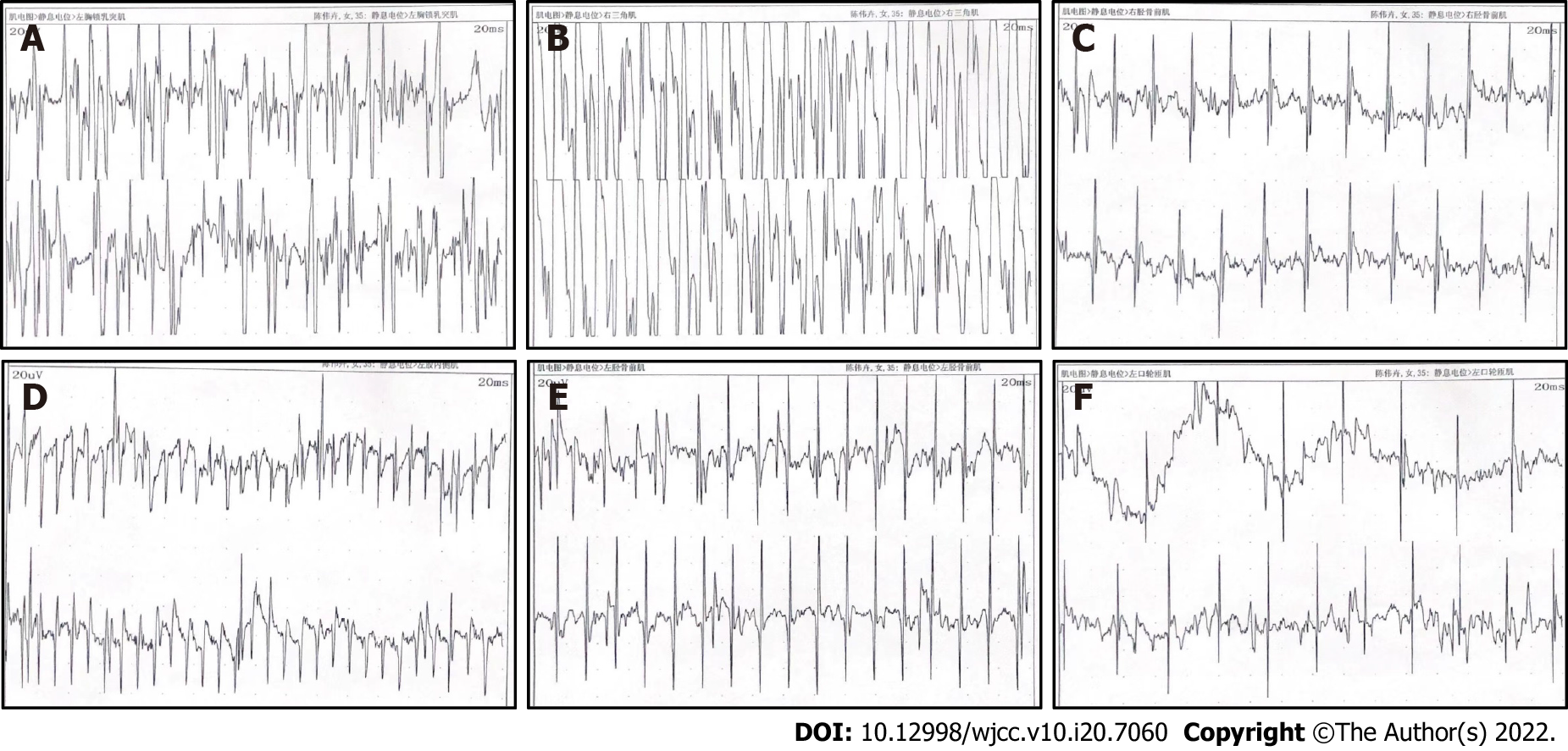
Figure 2 The patient's typical electromyography.
Electromyography of the proband: myotonia potentials were visible in the resting state of the examined extremity muscles and the orbicularis oris muscles, the motor unit potential time limit was shortened, and the amplitude was decreased. A: Left sternocleidomastoid muscle; B: Right deltoid; C: Right tibial anterior muscle; D: Left medial femoris muscle; E: Left tibial anterior muscle; F: Left orbicularis muscle.
- Citation: Jia YX, Dong CL, Xue JW, Duan XQ, Xu MY, Su XM, Li P. Myotonic dystrophy type 1 presenting with dyspnea: A case report. World J Clin Cases 2022; 10(20): 7060-7067
- URL: https://www.wjgnet.com/2307-8960/full/v10/i20/7060.htm
- DOI: https://dx.doi.org/10.12998/wjcc.v10.i20.7060


