Published online Jun 26, 2022. doi: 10.12998/wjcc.v10.i18.6136
Peer-review started: October 23, 2021
First decision: November 18, 2021
Revised: November 27, 2021
Accepted: April 21, 2022
Article in press: April 21, 2022
Published online: June 26, 2022
Processing time: 236 Days and 18.9 Hours
A right-sided sigmoid colon is an extremely rare anatomic variation that should be considered as a possibility by surgeons and radiologists before surgery. Here, we report the first clinical case of a carcinoma in a right-sided sigmoid colon revealed by a preoperative computed tomography (CT).
A 56-year-old Chinese man was admitted to the hospital with abdominal pain. CT revealed a redundant sigmoid colon with a mass on the right side of the cecum and ascending colon. Laparoscopy confirmed an abnormal course in the descending colon and sigmoid colon. Subsequently, hemicolectomy was performed in an open manner after laparoscopic exploration. Pathological examination revealed an infiltrative mucinous adenocarcinoma with two lymph node metastases. The patient was discharged without any complications after a week. There were no signs of recurrence or metastasis during the 3-month follow-up period.
We report a rare anomaly of a right-sided sigmoid colon with carcinoma, which should be differentiated from ascending colon cancer and pericecal hernia to prevent errors and other surgical complications.
Core Tip: The right-sided sigmoid colon was first described by a few cadaveric studies and may not have been fully recognized by clinicians in recent years due to its relative rarity. Recognizing this variation is essential for interventional and diagnostic colonoscopy and associated surgeries. This is the first clinical case of a carcinoma located in a right-sided sigmoid colon.
- Citation: Lyu LJ, Yao WW. Carcinoma located in a right-sided sigmoid colon: A case report. World J Clin Cases 2022; 10(18): 6136-6140
- URL: https://www.wjgnet.com/2307-8960/full/v10/i18/6136.htm
- DOI: https://dx.doi.org/10.12998/wjcc.v10.i18.6136
Recognizing variations in the sigmoid colon is of key importance to surgeons and radiologists. Here, we report an atypical anatomic variation of a right-sided sigmoid colon with carcinoma, which was incidentally observed during an emergency computed tomography (CT) scan for abdominal pain. To the best of our knowledge, this is the first clinical case of a carcinoma located in a right-sided sigmoid colon detected by a preoperative CT scan. We present the following case in accordance with the CARE Reporting Checklist.
A 56-year-old Chinese man was admitted to our hospital with abdominal pain in the right lower quadrant for 3 days.
The patient had experienced irregular and formless bowel movements for three months prior to presentation at our hospital. The patient also had right lower quadrant abdominal pain, which could not be relieved after defecation. The patient had no other accompanying symptoms, including any obvious symptoms of bowel obstruction.
The patient had no previous surgical history.
The patient did not have a history of smoking or drinking. There was no personal or family history of acute or chronic diseases.
The patient was 169 cm tall and weighed 65 kg. Physical examination revealed tenderness in the right lower quadrant, but no palpable mass was found.
Laboratory tests showed moderate increases in platelets and CA724, and normal levels of white blood cells and CA199. No abnormal results were found in other biochemical tests.
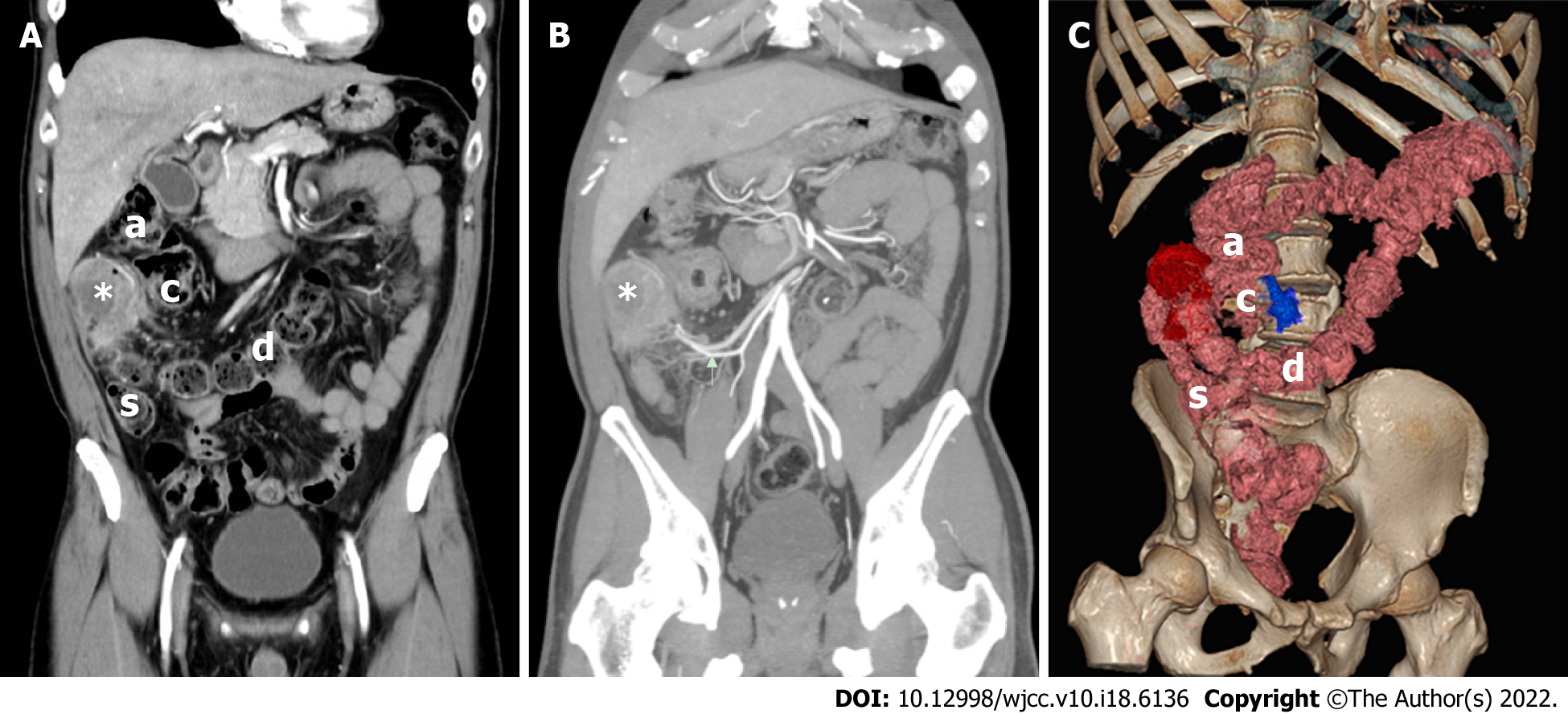
CT showed an atypical location of the redundant sigmoid colon with heterogeneously enhanced circumferential wall thickening on the right side of the cecum and ascending colon (Figures 1 and 2). We reported the atypical location of the sigmoid colon with a mass, suspected the presence of a pericecal hernia, and informed the surgeons accordingly.
Colonoscopy showed an annular stricture of the sigmoid colon caused by a tumor 28 cm from the anus. Subsequent biopsy indicated malignancy.
Due to inconclusive radiological signs, the patient underwent laparoscopic exploration. Intraoperatively, the surgeons detected atypical positions of both the sigmoid colon and descending colon. They converted the surgery to an open operation for safety considerations.
The surgeons found a mass that almost completely blocked the lumen of the sigmoid colon. The redundant sigmoid colon was located on the right side of the ascending colon and cecum and was supplied by the branches of the inferior mesenteric artery, which ran to the right instead of its standard left-sided course. The descending colon started at the splenic flexure and crossed to the right at the level of the L4 vertebra, and occupied the subhepatic region to continue as the sigmoid colon. The small intestine was normal. Pathological examination revealed an infiltrative mucinous adenocarcinoma with two lymph node metastases, pT4N1M0 stage IIIB (UICC-TNM: 8th edition).
The surgeons performed a hemicolectomy with regional lymphadenectomy during laparotomy. No bowel perforation was observed.
The patient was discharged on postoperative day 7 without any complications. Moreover, there were no signs of recurrence or metastasis during the 3-month follow-up period.
The sigmoid colon shows the greatest variation in length and position[1]. The variation in length is mainly related to racial differences and a high-fiber diet[2], the position of the sigmoid colon loop is a means of adapting to the general length of the sigmoid colon[2]. A study by Saxena et al[3] suggested that the sigmoid colon of young children (age < 5 years) is often situated entirely on the right side for redundancy; this is not the case in adults. The presence of a right-sided sigmoid colon is very rare in adults and may be related to fixation anomalies[1], redundancy of the colon, or secondary rotation of the colon during embryogenesis[4].
In most cases of right-side colon carcinoma, the carcinoma is observed to be located in the ascending colon. In our case, circumferential wall thickening of the colon occupied the subhepatic region on the right of the ascending colon, and the ileocecal junction was displaced toward the left at the level of the L4 transverse process instead of the right pelvic region, indicating that the cecum was undescended due to midgut malrotation during embryogenesis[4]. Colonoscopy revealed a stricture 28 cm from the anus, demonstrating a sigmoid colon other than the ascending colon. The identification of the tumor location is crucial for determining the appropriate clinical course because the sigmoid colon and ascending colon have different embryological origins[5], and a recent study[6] demonstrated that right-sided colon carcinomas exhibit exophytic pathological behavior and poorer overall survival than left-sided colon carcinomas.
In the present case, the colon with wall thickening on the right side of the ascending colon was similar to a rare type of internal hernia occurring near the cecum, namely, pericecal hernia[7]. However, pericecal hernia usually involves a small bowel other than the sigmoid colon; such pericecal hernias usually produce acute intestinal obstruction, which can be confirmed by CT. In addition, it is expected that the descending colon and sigmoid colon should be observed in the standard position relative to the pericecal hernia and the inferior mesenteric artery. On CT images of the present case, no small bowel herniation or obvious dilation was observed. The descending colon crossed to the right side at the level of the L4 vertebra, where it entered the peritoneal cavity and continued as the sigmoid colon on the right side. Notably, the inferior mesenteric artery ran to the right instead of its normal left-sided course.
Shrivastava et al[8] first described the right-sided sigmoid colon in a cadaveric study in 2013. Flores-Ríos et al[9] reported a case of secondary right-sided descending and sigmoid colon caused by a wandering spleen due to laxity or abnormal development of the peritoneal ligaments, which was different from our case. Subsequently, there were two case reports[1,10] of right-sided sigmoid colons, which were discovered incidentally during surgery.
To the best of our knowledge, this is the first clinical case of carcinoma located in the right-sided sigmoid colon revealed by a preoperative CT scan and confirmed by surgery. Surgeons and radiologists should be aware of this rare variation when examining patients experiencing abdominal pain in the right lower quadrant.
The limitation of our case was the lack of appropriate intraoperative images compatible with the volume rendering image (Figure 2C), which could have provided readers with an intuitive understanding.
We report a rare anomaly of the right-sided sigmoid colon with carcinoma that could be detected by careful examination with a preoperative CT scan. This is a major congenital colon anomaly that should be recognized preoperatively and needs to be differentiated from the ascending colon and pericecal hernia to prevent errors and other surgical complications.
Provenance and peer review: Unsolicited article; Externally peer reviewed.
Peer-review model: Single blind
Specialty type: Anatomy and morphology
Country/Territory of origin: China
Peer-review report’s scientific quality classification
Grade A (Excellent): 0
Grade B (Very good): B, B
Grade C (Good): 0
Grade D (Fair): 0
Grade E (Poor): E
P-Reviewer: Ding X, China; Haddadi S, Spain; Oley MH, Indonesia S-Editor: Ma YJ L-Editor: A P-Editor: Ma YJ
| 1. | Kar H, Karasu S, Bag H, Cin N, Durak E, Peker Y, Tatar FA. Case Report of the Descending Colon Located Completely in the Right Extraperitoneal Cavity and Sigmoid Colon Located in the Right Quadrant in a Patient with a Splenic Flexure Tumor: a Rare Anomaly. Indian J Surg. 2016;78:323-325. [RCA] [PubMed] [DOI] [Full Text] [Cited by in Crossref: 1] [Cited by in RCA: 1] [Article Influence: 0.1] [Reference Citation Analysis (1)] |
| 2. | Madiba TE, Haffajee MR. Sigmoid colon morphology in the population groups of Durban, South Africa, with special reference to sigmoid volvulus. Clin Anat. 2011;24:441-453. [RCA] [PubMed] [DOI] [Full Text] [Cited by in Crossref: 18] [Cited by in RCA: 21] [Article Influence: 1.5] [Reference Citation Analysis (0)] |
| 3. | Saxena AK, Sodhi KS, Tirumani S, Mumtaz HA, Narasimha Rao KL, Khandelwal N. Position of a sigmoid colon in right iliac fossa in children: A retrospective study. J Indian Assoc Pediatr Surg. 2011;16:93-96. [RCA] [PubMed] [DOI] [Full Text] [Cited by in Crossref: 3] [Cited by in RCA: 3] [Article Influence: 0.2] [Reference Citation Analysis (0)] |
| 4. | Gnansekaran D, Prashant SA, Veeramani R, Yekappa SH. Congenital positional anomaly of descending colon and sigmoid colon: Its embryological basis and clinical implications. Med J Armed Forces India. 2021;77:241-244. [RCA] [PubMed] [DOI] [Full Text] [Cited by in Crossref: 1] [Cited by in RCA: 1] [Article Influence: 0.3] [Reference Citation Analysis (1)] |
| 5. | Jess P, Hansen IO, Gamborg M, Jess T; Danish Colorectal Cancer Group. A nationwide Danish cohort study challenging the categorisation into right-sided and left-sided colon cancer. BMJ Open. 2013;3. [RCA] [PubMed] [DOI] [Full Text] [Full Text (PDF)] [Cited by in Crossref: 58] [Cited by in RCA: 78] [Article Influence: 6.5] [Reference Citation Analysis (0)] |
| 6. | Xie X, Zhou Z, Song Y, Wang W, Dang C, Zhang H. Differences between carcinoma of the cecum and ascending colon: Evidence based on clinical and embryological data. Int J Oncol. 2018;53:87-98. [RCA] [PubMed] [DOI] [Full Text] [Full Text (PDF)] [Cited by in Crossref: 3] [Cited by in RCA: 8] [Article Influence: 1.1] [Reference Citation Analysis (0)] |
| 7. | Inukai K, Tsuji E, Uehara S. Paracecal hernia with intestinal ischemia treated with laparoscopic assisted surgery. Int J Surg Case Rep. 2018;44:20-23. [RCA] [PubMed] [DOI] [Full Text] [Full Text (PDF)] [Cited by in Crossref: 12] [Cited by in RCA: 11] [Article Influence: 1.6] [Reference Citation Analysis (0)] |
| 8. | Shrivastava P, Tuli A, Kaur S, Raheja S. Right sided descending and sigmoid colon: its embryological basis and clinical implications. Anat Cell Biol. 2013;46:299-302. [RCA] [PubMed] [DOI] [Full Text] [Full Text (PDF)] [Cited by in Crossref: 8] [Cited by in RCA: 10] [Article Influence: 0.8] [Reference Citation Analysis (0)] |
| 9. | Flores-Ríos E, Méndez-Díaz C, Rodríguez-García E, Pérez-Ramos T. Wandering spleen, gastric and pancreatic volvulus and right-sided descending and sigmoid colon. J Radiol Case Rep. 2015;9:18-25. [RCA] [PubMed] [DOI] [Full Text] [Cited by in Crossref: 4] [Cited by in RCA: 13] [Article Influence: 1.3] [Reference Citation Analysis (0)] |
| 10. | Watanabe K, Abiko K, Tamura S, Katsumata M, Amano Y, Takao Y. Right-sided Sigmoid Colon Revealed during Laparoscopic Sacrocolpopexy. J Minim Invasive Gynecol. 2021;28:1267-1268. [RCA] [PubMed] [DOI] [Full Text] [Cited by in Crossref: 1] [Cited by in RCA: 4] [Article Influence: 0.8] [Reference Citation Analysis (0)] |


