Copyright
©The Author(s) 2022.
World J Clin Cases. Jun 16, 2022; 10(17): 5846-5853
Published online Jun 16, 2022. doi: 10.12998/wjcc.v10.i17.5846
Published online Jun 16, 2022. doi: 10.12998/wjcc.v10.i17.5846
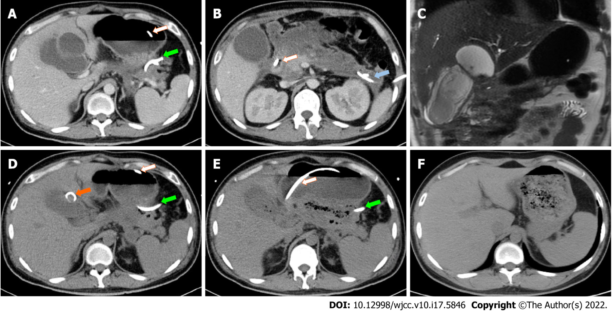
Figure 2 Gallbladder perforation and cholecysto-colonic fistula during the course of disease.
The jejunal feeding tube is marked by a white arrow. The percutaneous drainage tube for the pancreatic tail area is marked by a green arrow. The percutaneous drainage tube for the left paracolic sulcus is marked by a blue arrow. A: Contrast-enhanced computed tomography (CT) demonstrating a cystic lesion communicating to the gallbladder 1 mo after severe acute pancreatitis (SAP) onset; B: Contrast-enhanced CT demonstrating a large gallbladder and profound exudation in pancreatic head region 1 mo after SAP onset; C: Magnetic resonance cholangiopancreatography demonstrating a cystic lesion adjacent to the gallbladder 1 mo after SAP onset; D: CT demonstrating adequate drainage of gallbladder perforation 2 mo after SAP onset. The percutaneous drainage tube for the cystic lesion is marked by an orange arrow; E: CT demonstrating gas in the gallbladder lumen as indirect evidence of cholecysto-colonic fistula before debridement surgery; F: CT demonstrating a recovery from cholecystectomy 10 mo after SAP onset.
- Citation: Wang QP, Chen YJ, Sun MX, Dai JY, Cao J, Xu Q, Zhang GN, Zhang SY. Spontaneous gallbladder perforation and colon fistula in hypertriglyceridemia-related severe acute pancreatitis: A case report. World J Clin Cases 2022; 10(17): 5846-5853
- URL: https://www.wjgnet.com/2307-8960/full/v10/i17/5846.htm
- DOI: https://dx.doi.org/10.12998/wjcc.v10.i17.5846


