Copyright
©The Author(s) 2022.
World J Clin Cases. Jun 16, 2022; 10(17): 5748-5755
Published online Jun 16, 2022. doi: 10.12998/wjcc.v10.i17.5748
Published online Jun 16, 2022. doi: 10.12998/wjcc.v10.i17.5748
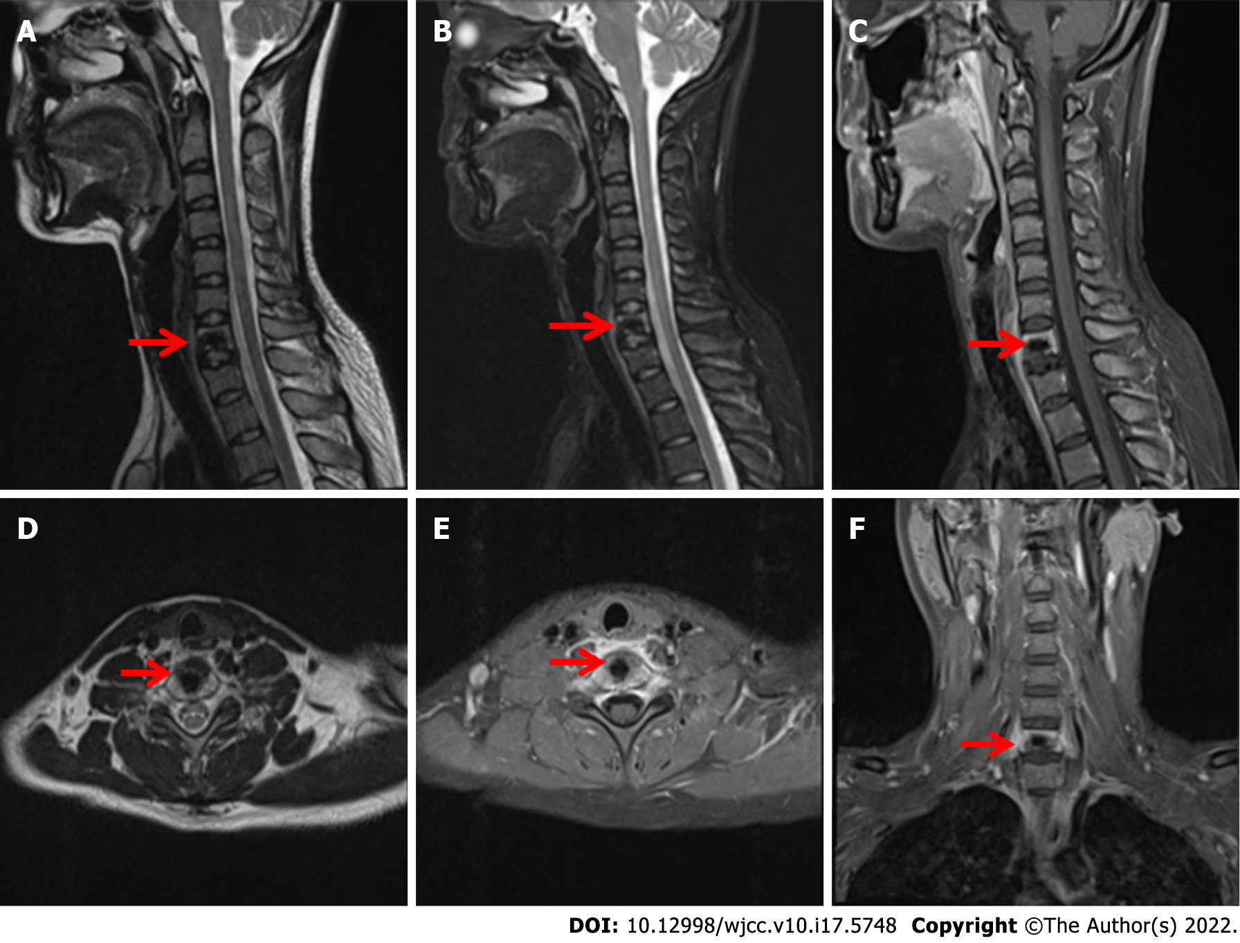
Figure 2 Magnetic resonance imaging of the cervical vertebra shows a patchy, abnormal signal shadow of the C7 vertebral body, swelling of adjacent soft tissue, uneven enhancement after enhancement, and no abnormal signal of the cervical spinal cord.
A, D: T1-weighted images; B: T2-weighted images; C, E and F: Fat suppression images.
- Citation: Li C, Li S, Hu W. Chondromyxoid fibroma of the cervical spine: A case report. World J Clin Cases 2022; 10(17): 5748-5755
- URL: https://www.wjgnet.com/2307-8960/full/v10/i17/5748.htm
- DOI: https://dx.doi.org/10.12998/wjcc.v10.i17.5748


