Copyright
©The Author(s) 2022.
World J Clin Cases. Jun 16, 2022; 10(17): 5717-5722
Published online Jun 16, 2022. doi: 10.12998/wjcc.v10.i17.5717
Published online Jun 16, 2022. doi: 10.12998/wjcc.v10.i17.5717
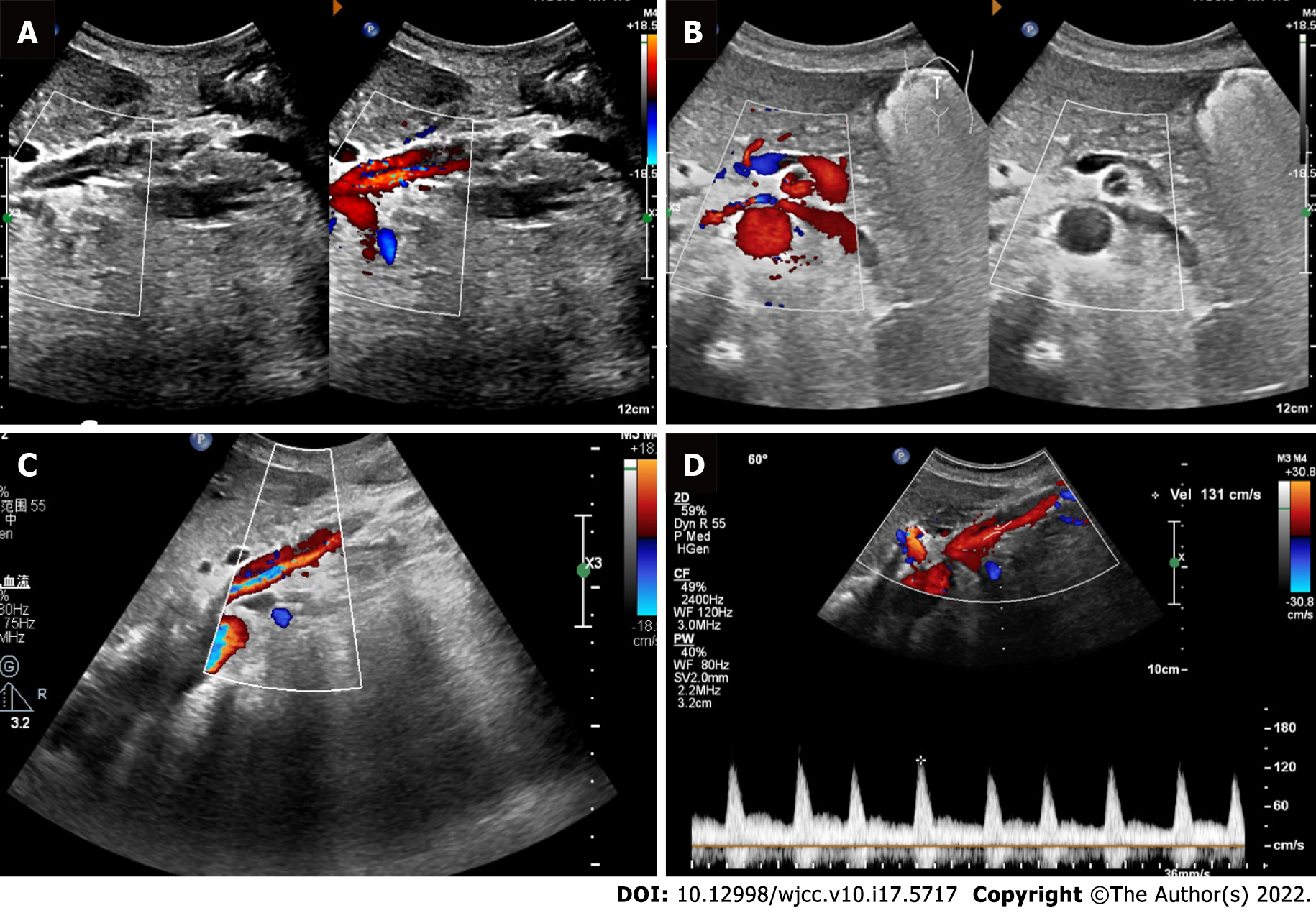
Figure 2 Doppler ultrasound showed the blood flow of the superior mesenteric artery dissection.
A: Ultrasonic longitudinal view showed the flow at the opening of the superior mesenteric artery dissection (SISMAD); B: Ultrasonic transverse view showed the flow at the opening of the SISMAD; C: Color Doppler flow imaging showed the true and false lumens of the SISMAD; D: True lumen velocity of superior mesenteric artery dissection was measured by spectral Doppler.
- Citation: Zhang Y, Zhou JY, Liu J, Bai C. Diagnosis of spontaneous isolated superior mesenteric artery dissection with ultrasound: A case report. World J Clin Cases 2022; 10(17): 5717-5722
- URL: https://www.wjgnet.com/2307-8960/full/v10/i17/5717.htm
- DOI: https://dx.doi.org/10.12998/wjcc.v10.i17.5717


