Published online Apr 16, 2013. doi: 10.12998/wjcc.v1.i1.28
Revised: December 18, 2012
Accepted: January 29, 2013
Published online: April 16, 2013
Processing time: 165 Days and 13.5 Hours
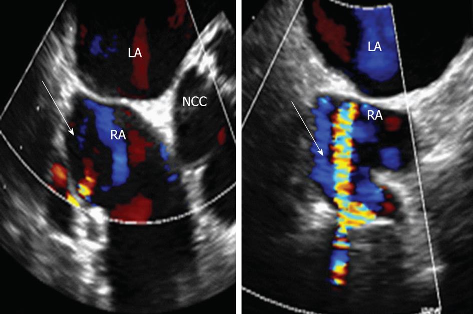
An unknown aberrant flow in the right atrium observed on doppler with transesophageal echocardiogram (TEE) in a patient with prior coronary bypass. TEE revealed normal size left ventricle with severely dilated left atrium. There was moderate aortic regurgitation and moderate aortic stenosis noted. Patient was incidentally found to have an abnormal vascular communication noted to the right atrium. To further evaluate this finding, the patient underwent cardiac magnetic resonance angiography which revealed that the tubular structure noted on TEE was actually a graft that was abutting onto the coronary sinus, and the flow anomaly was really the graft coming up and running adjacent to the coronary sinus.
- Citation: Cao L, Farooqui MA, Wood W, Cahill J, Movahed A. Saphenous graft on transesophageal echocardiogram masquerading as an abnormal vascular communication into the right atrium. World J Clin Cases 2013; 1(1): 28-30
- URL: https://www.wjgnet.com/2307-8960/full/v1/i1/28.htm
- DOI: https://dx.doi.org/10.12998/wjcc.v1.i1.28
Cardiovascular disease is the leading cause of death in the United States. The number is expected to increase substantially. As a result, significant advancements have been made to diagnosed, treat and also to prevent this epidemic. More than 150 000 people undergo coronary artery bypass grafting each year in the United States[1-4]. Many of these patients will have routine follow ups and among them, several will have subsequent imaging studies to evaluate improvement, or unfortunately, disease progression. Echocardiography is and has been a great tool for the evaluation of cardiac anatomy and function. Postoperative patients for valve replacements and or coronary bypass grafts have offered interesting and challenging studies that often show the limitations of echocardiography[1,5]. Magnetic resonance imaging (MRI) is a non-invasive modality which can be used for direct visualization of coronary artery bypass grafts. Along with spin-echo and gradient-echo (cine-MRI), they provide information on graft morphology and patency with a 90% accuracy[3,6,7].
We present a case of a 72-year-old male with past medical history of moderate aortic stenosis, coronary artery disease status post five vessel coronary artery bypass grafting with left internal mammary artery to left anterior descending, saphenous vein graft to the third obtuse marginal branch, saphenous vein graft to right coronary artery, saphenous vein graft to first diagonal, and saphenous vein graft to ramus intermedius. Patient was evaluated at the cardiology clinic for further stratification of aortic stenosis and underwent left atrium transesophageal echocardiogram (TEE). TEE revealed normal size left ventricle with severely dilated left atrium. There was moderate aortic regurgitation and moderate aortic stenosis noted. Patient was incidentally found to have an abnormal vascular communication noted to the right atrium (Figure 1). To further evaluate this finding, the patient underwent cardiac magnetic resonance angiography (MRA) which revealed that the tubular structure noted on TEE was actually a graft that was abutting onto the coronary sinus, and the flow anomaly was really the graft coming up and running adjacent to the coronary sinus (Figure 2).
This case shows one way a graft can appear on TEE, which was confusing until MRA and clinical correlation were accomplished. This case also illustrates the importance of correlating graft locations when performing TEE and also to utilize cardiac MRA if needed to better define unusual presentations of sapheno-venous graft adjacent to atrial chambers.
P- Reviewer Vermeersch P S- Editor Zhai HH L- Editor A E- Editor Zheng XM
| 1. | Khabeishvili G, Shaburishvili T, Wann S, Sampson C, Manley JC. Saphenous vein graft pseudoaneurysm: diagnosis by transesophageal echocardiography and magnetic resonance imaging. J Am Soc Echocardiogr. 1995;8:338-340. [RCA] [PubMed] [DOI] [Full Text] [Cited by in Crossref: 9] [Cited by in RCA: 10] [Article Influence: 0.3] [Reference Citation Analysis (0)] |
| 2. | Le Breton H, Pavin D, Langanay T, Roland Y, Leclercq C, Beliard JM, Bedossa M, Rioux C, Pony JC. Aneurysms and pseudoaneurysms of saphenous vein coronary artery bypass grafts. Heart. 1998;79:505-508. [PubMed] |
| 3. | Memon AQ, Huang RI, Marcus F, Xavier L, Alpert J. Saphenous vein graft aneurysm: case report and review. Cardiol Rev. 2003;11:26-34. [RCA] [PubMed] [DOI] [Full Text] [Cited by in Crossref: 42] [Cited by in RCA: 43] [Article Influence: 2.0] [Reference Citation Analysis (0)] |
| 4. | Wight JN, Salem D, Vannan MA, Pandian NG, Rozansky MI, Dohan MC, Rastegar H. Asymptomatic large coronary artery saphenous vein bypass graft aneurysm: a case report and review of the literature. Am Heart J. 1997;133:454-460. [RCA] [PubMed] [DOI] [Full Text] [Cited by in Crossref: 29] [Cited by in RCA: 30] [Article Influence: 1.1] [Reference Citation Analysis (0)] |
| 5. | Bansal RC. Echocardiographic diagnosis of an asymptomatic aneurysm of a saphenous vein graft. J Am Soc Echocardiogr. 2002;15:661-664. [RCA] [PubMed] [DOI] [Full Text] [Cited by in Crossref: 4] [Cited by in RCA: 5] [Article Influence: 0.2] [Reference Citation Analysis (1)] |
| 6. | Yohann MM, Gilkeson RC, Markowitz AH, el-Zein C. Coronary artery graft aneurysm diagnosed by cardiac magnetic resonance imaging. Ann Thorac Surg. 2000;70:1729. [PubMed] |
| 7. | Sharifi M, Dillon JC, Pompili VJ. Saphenous vein graft ectasia: an unusual late complication of coronary artery bypass surgery. A case report. Angiology. 1999;50:497-501. [RCA] [PubMed] [DOI] [Full Text] [Cited by in Crossref: 6] [Cited by in RCA: 8] [Article Influence: 0.3] [Reference Citation Analysis (0)] |


