Copyright
©The Author(s) 2021.
World J Clin Cases. Mar 6, 2021; 9(7): 1741-1747
Published online Mar 6, 2021. doi: 10.12998/wjcc.v9.i7.1741
Published online Mar 6, 2021. doi: 10.12998/wjcc.v9.i7.1741
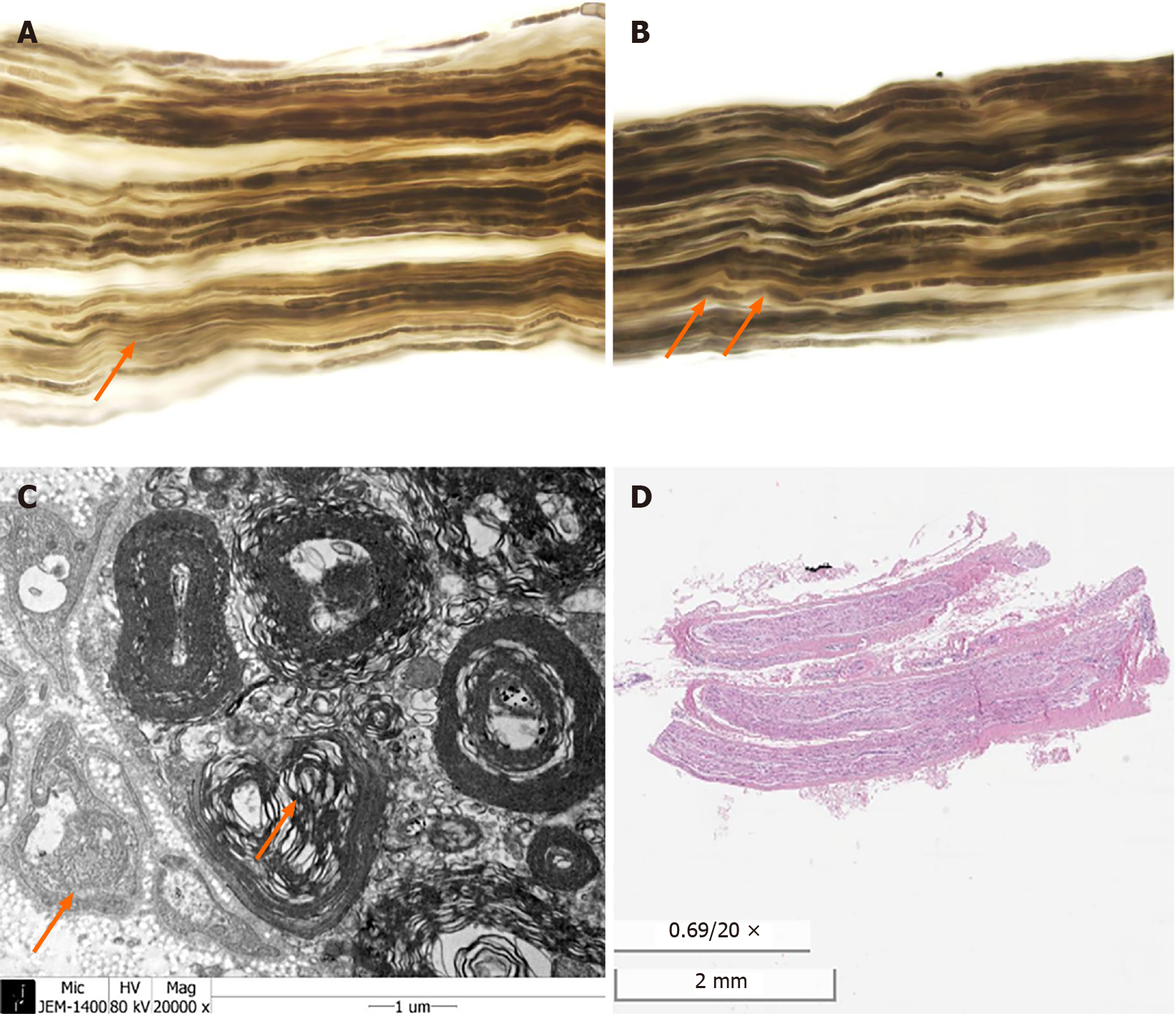
Figure 1 Sural nerve biopsy.
A: Nerve teasing. Decreased number of nerve fibers; B: Nerve teasing. Breakdown of myelin sheath; C: Electron microscopy. Degeneration of myelinated nerve fiber; D: Hematoxylin and eosin stain. No definite inflammatory cell infiltration or vasculitis.
- Citation: Chae HJ, Kim JW, Lee YL, Park JH, Lee SY. Mononeuropathy multiplex associated with systemic vasculitis: A case report. World J Clin Cases 2021; 9(7): 1741-1747
- URL: https://www.wjgnet.com/2307-8960/full/v9/i7/1741.htm
- DOI: https://dx.doi.org/10.12998/wjcc.v9.i7.1741


