Copyright
©The Author(s) 2021.
World J Clin Cases. Mar 6, 2021; 9(7): 1728-1733
Published online Mar 6, 2021. doi: 10.12998/wjcc.v9.i7.1728
Published online Mar 6, 2021. doi: 10.12998/wjcc.v9.i7.1728
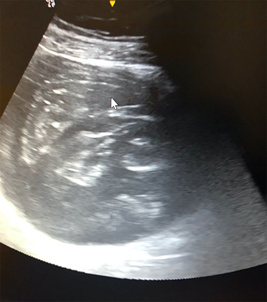
Figure 1 Ultrasound examination showing a voluminous septate cystic mass.
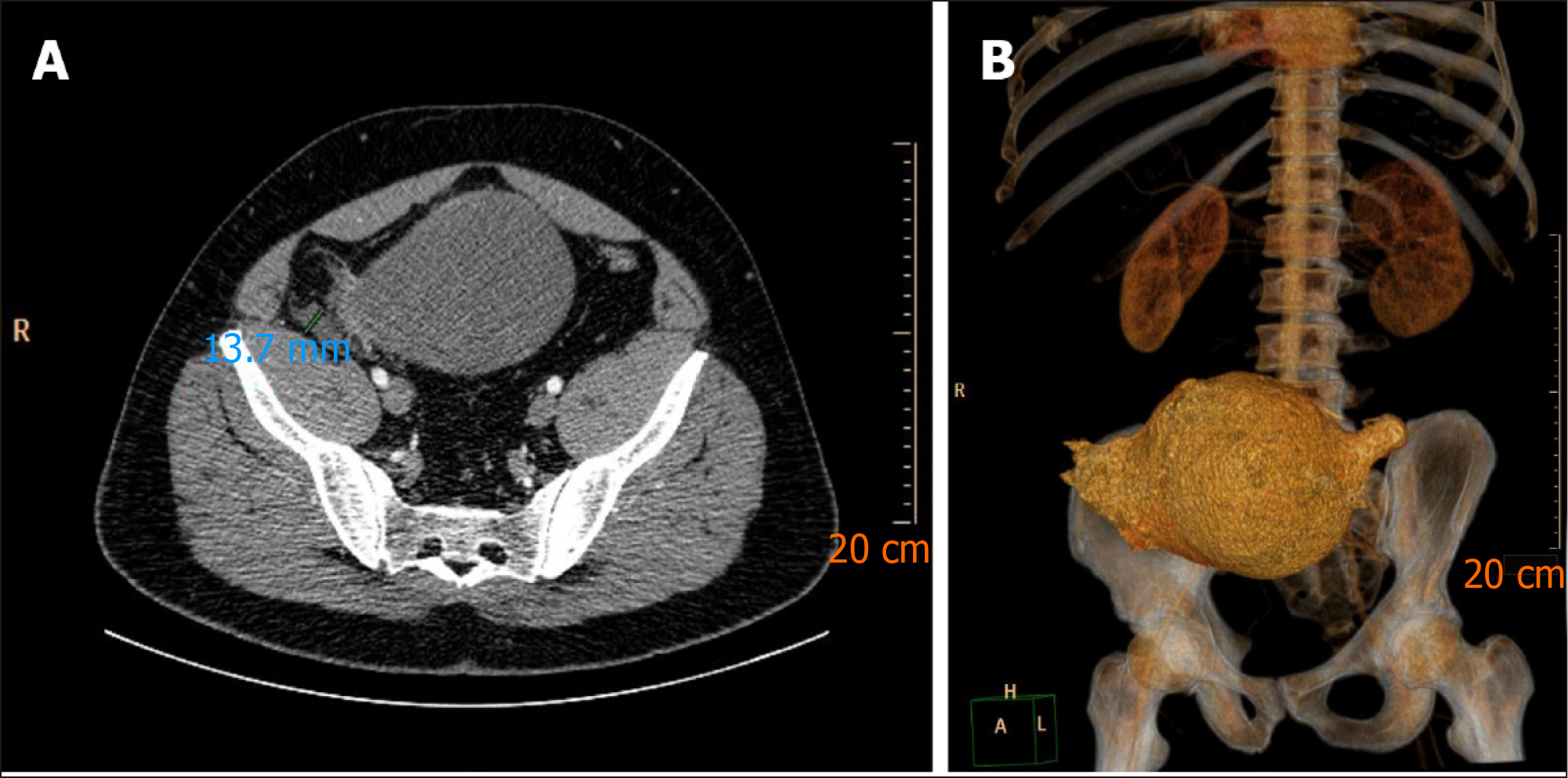
Figure 2 Computed tomography findings.
A: Computed tomography (CT) scan showing voluminous cystic tumor communicating with the cecum; B: CT scan showing reconstruction image of the same lesion.
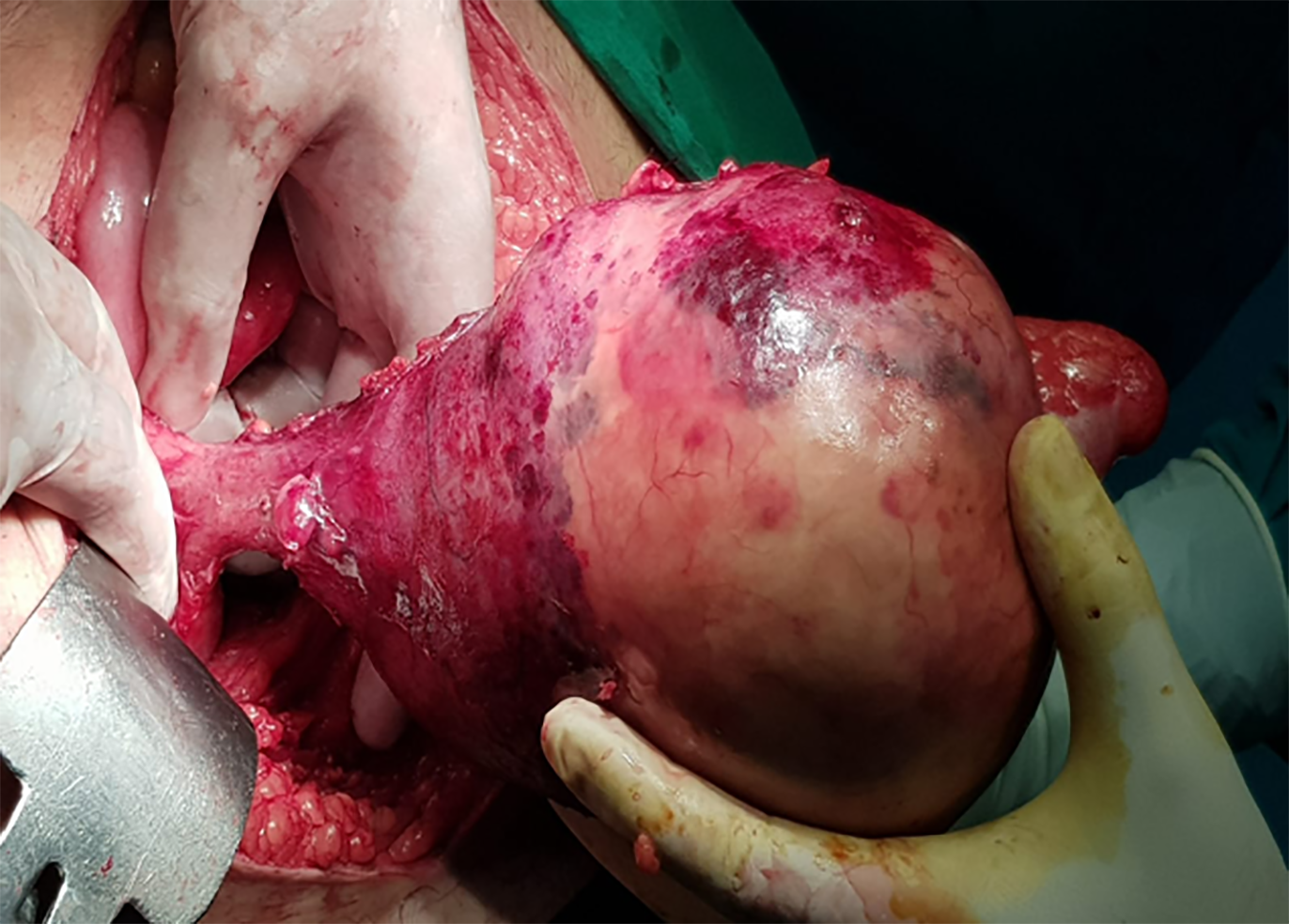
Figure 3 Intra-operatory view of a large tumor implanted in the cecum.
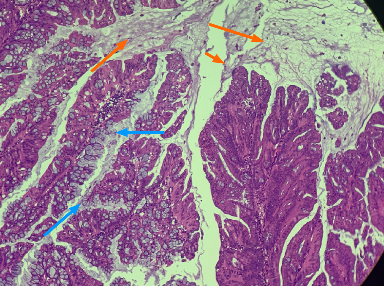
Figure 4 Hematoxylin and eosin stain (40 ×) depicting villous mucus secreting dysplastic epithelium (blue arrows) with dissecting pools of mucin (orange arrows).
- Citation: Chirca A, Negreanu L, Iliesiu A, Costea R. Mucinous appendiceal neoplasm: A case report. World J Clin Cases 2021; 9(7): 1728-1733
- URL: https://www.wjgnet.com/2307-8960/full/v9/i7/1728.htm
- DOI: https://dx.doi.org/10.12998/wjcc.v9.i7.1728


