Copyright
©The Author(s) 2021.
World J Clin Cases. Mar 6, 2021; 9(7): 1705-1713
Published online Mar 6, 2021. doi: 10.12998/wjcc.v9.i7.1705
Published online Mar 6, 2021. doi: 10.12998/wjcc.v9.i7.1705
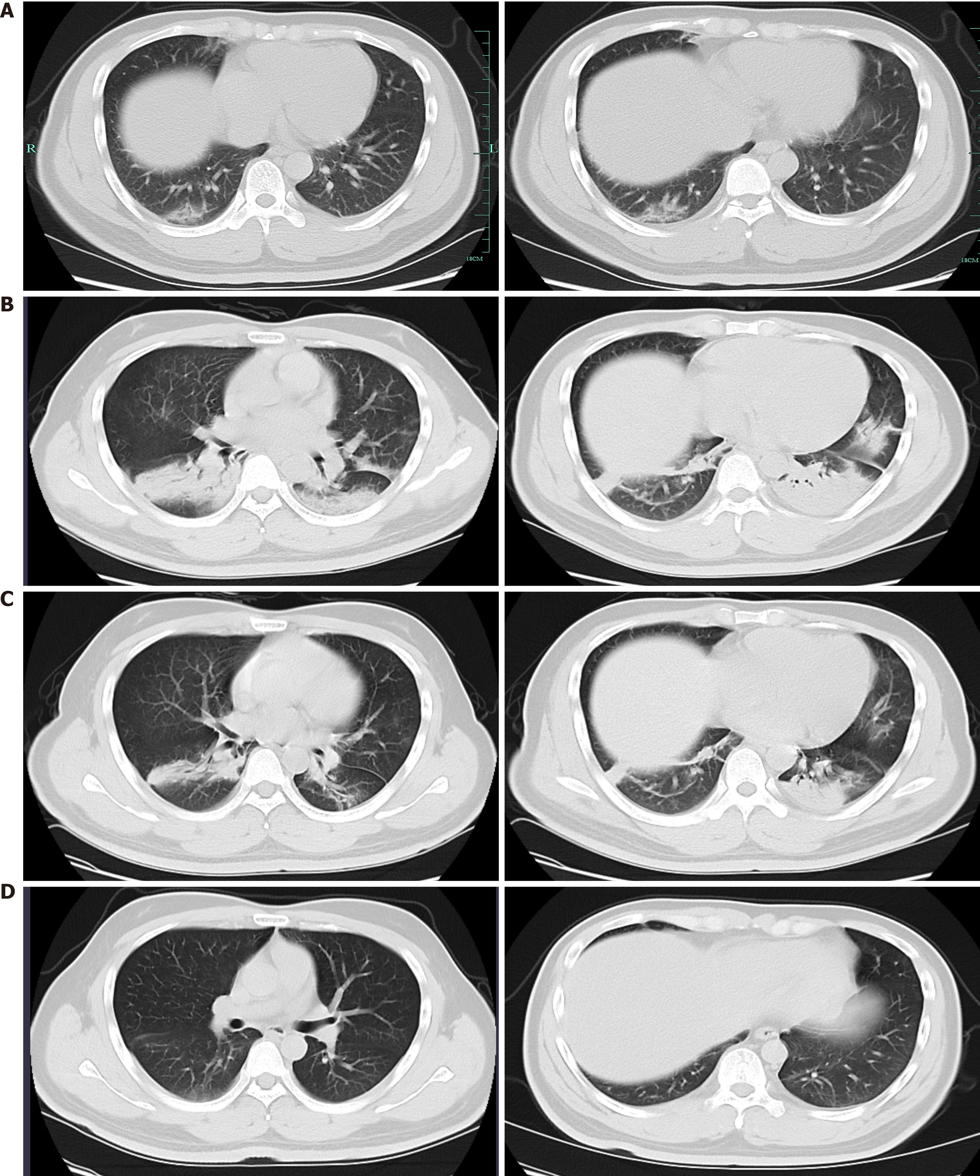
Figure 1 Chest computed tomography images of the patient with coronavirus disease 2019 in different stages of illness.
A: Interstitial changes in both lungs and ground-glass opacities (GGOs) in the subpleural area of the right lower lobe on January 19 (Illness day 1); B: Worsening basilar streaky opacities, patchy consolidations, and GGOs in both lungs on January 30 (Illness day 12); C: Significant absorption of patches infiltrating in both lungs on February 4 (Illness day 17); D: Almost no abnormality with a few fibrotic changes left in both lungs on February 19.
- Citation: Lu J, Xie ZY, Zhu DH, Li LJ. Human menstrual blood-derived stem cells as immunoregulatory therapy in COVID-19: A case report and review of the literature. World J Clin Cases 2021; 9(7): 1705-1713
- URL: https://www.wjgnet.com/2307-8960/full/v9/i7/1705.htm
- DOI: https://dx.doi.org/10.12998/wjcc.v9.i7.1705


