Copyright
©The Author(s) 2021.
World J Clin Cases. Mar 6, 2021; 9(7): 1668-1675
Published online Mar 6, 2021. doi: 10.12998/wjcc.v9.i7.1668
Published online Mar 6, 2021. doi: 10.12998/wjcc.v9.i7.1668
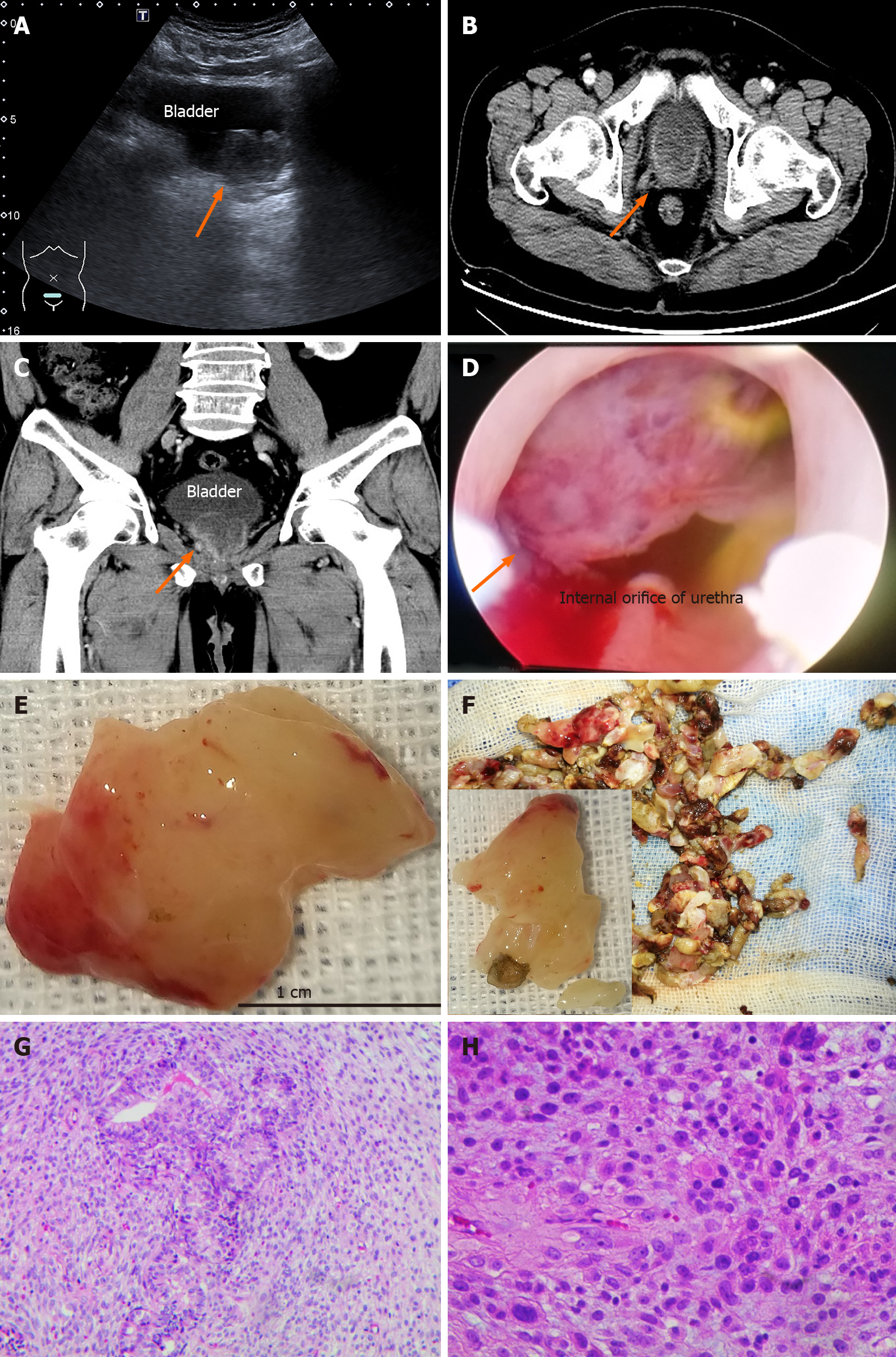
Figure 1 Clinical and pathological features of case 2 at the diagnosis of sarcomatoid carcinoma.
A: Ultrasound: The size of the prostate was 3.9 cm × 3.2 cm, and a 1.5 cm × 1.1 cm round mass was present in the gland (arrow); B and C: The mass was approximately 4.1 cm × 3.0 cm × 4.0 cm on computed tomography (arrow), and enhanced scanning was uneven. No obvious abnormality was found in the bilateral seminal vesicles; D-F: A white, narrow pedicled, spherical solid tumor blocked the internal orifice of the urethra. The spherical lesion mainly arose from the 8-11 o'clock position of the prostate and resembled fish flesh in sections (arrow); G and H: Various heteromorphic tumor cells showed infiltrating growth, which included immature small round cells, subepithelial cells, spindle cells, lipoblasts and tumor giant cells (arrow), etc. G: Original magnification × 100; H: × 200.
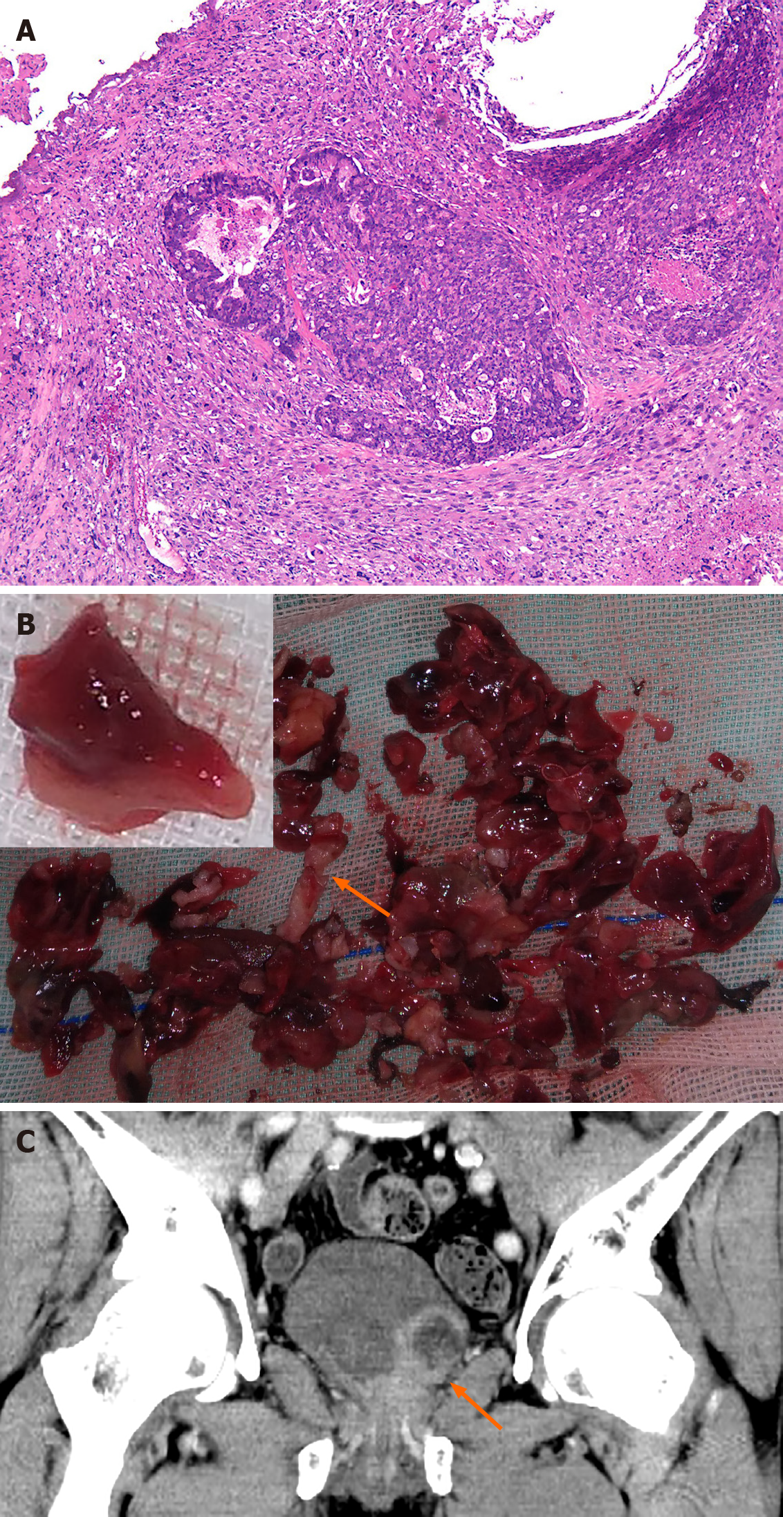
Figure 2 Clinical and pathological features of case 1 when diagnosed with sarcomatoid carcinoma.
A: Histopathology specimen showed poorly differentiated carcinoma with extensive necrosis. Adenocarcinoma involved < 2% of the prostate; B: Completely different lesions were dull red in color resembling fresh clots and were isolated and soft (resembling liver) in texture (orange arrow). However, the prostate cancer tissues were pale due to ischemia, as shown in the figure (orange arrow); C: Computed tomography scan showing an irregular mixed density range approximately 4.7 cm × 4.8 cm × 4.2 cm near the previous operation area (arrow). The mass extended from the prostate to the lateral wall of the bladder and the internal orifice of the urethra.
- Citation: Wei W, Li QG, Long X, Hu GH, He HJ, Huang YB, Yi XL. Sarcomatoid carcinoma of the prostate with bladder invasion shortly after androgen deprivation: Two case reports. World J Clin Cases 2021; 9(7): 1668-1675
- URL: https://www.wjgnet.com/2307-8960/full/v9/i7/1668.htm
- DOI: https://dx.doi.org/10.12998/wjcc.v9.i7.1668


