Copyright
©The Author(s) 2021.
World J Clin Cases. Mar 6, 2021; 9(7): 1639-1645
Published online Mar 6, 2021. doi: 10.12998/wjcc.v9.i7.1639
Published online Mar 6, 2021. doi: 10.12998/wjcc.v9.i7.1639
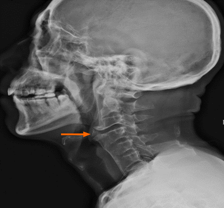
Figure 1 Patient's preoperative cervical spine lateral X-ray examination showing prominent osteophyte formation anteriorly on the left at the C3-4 level.
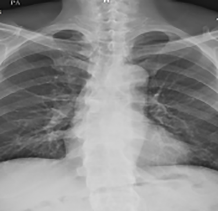
Figure 2 Simple thoracolumbar radiography showing a bamboo spine appearance.
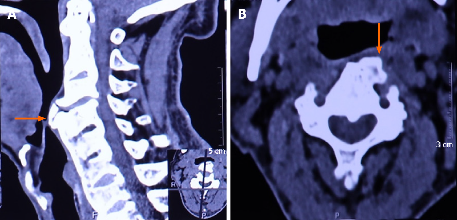
Figure 3 Patient's preoperative three-dimensional computed tomography examinations of cervical spine.
A: Sagittal plane; B: Coronal plane.
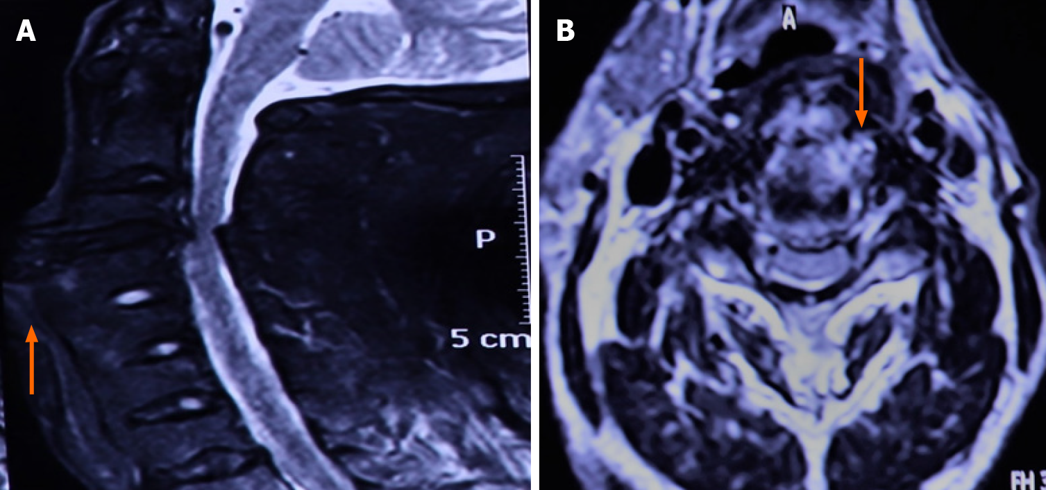
Figure 4 Patient's preoperative magnetic resonance imaging examinations of cervical spine.
A: Sagittal plane; B: Coronal plane.
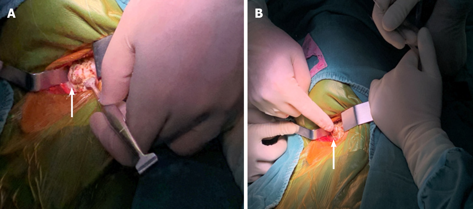
Figure 5 Pictures during the surgery.
A and B: Giant anterior cervical osteophyte.
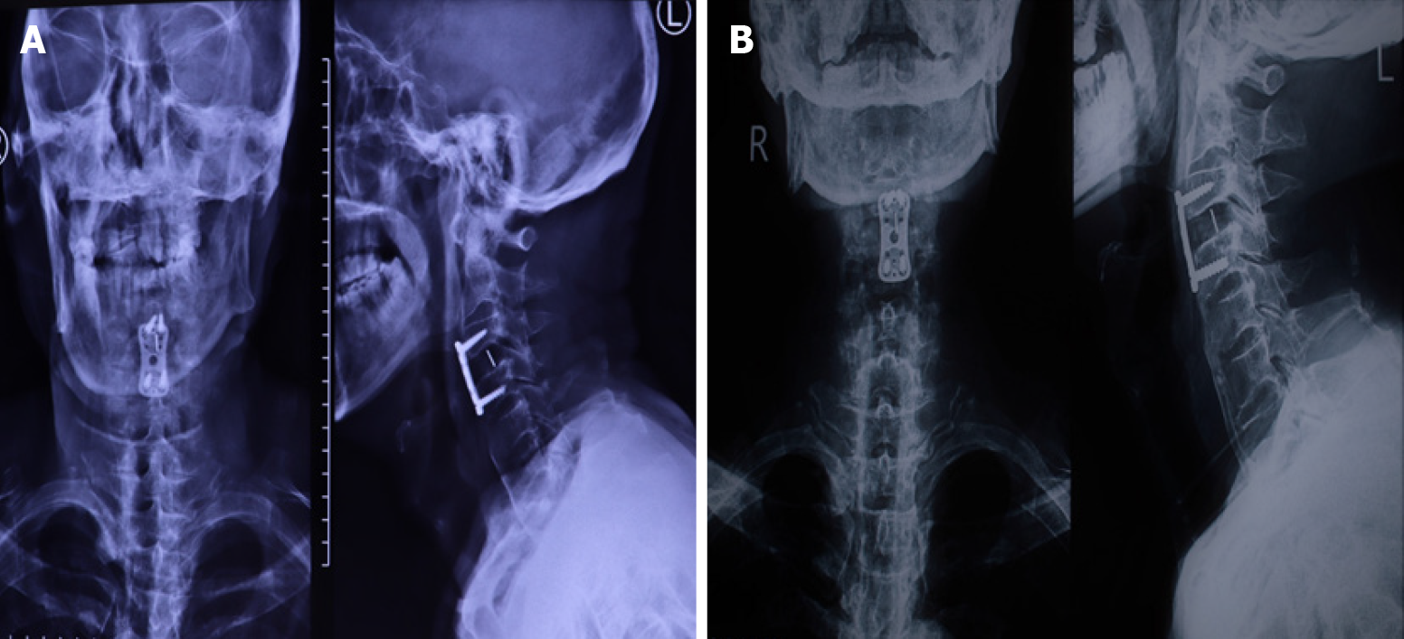
Figure 6 Patient's postoperative X-ray examinations.
A: Lateral radiograph of cervical spine 1 d after operation; B: At 1 mo after operation, indicating the follow-up of patients in different time periods after surgery.
- Citation: Wang XW, Zhang WZ. Dysphagia in a patient with ankylosing spondylitis: A case report. World J Clin Cases 2021; 9(7): 1639-1645
- URL: https://www.wjgnet.com/2307-8960/full/v9/i7/1639.htm
- DOI: https://dx.doi.org/10.12998/wjcc.v9.i7.1639


