Copyright
©The Author(s) 2021.
World J Clin Cases. Feb 26, 2021; 9(6): 1490-1498
Published online Feb 26, 2021. doi: 10.12998/wjcc.v9.i6.1490
Published online Feb 26, 2021. doi: 10.12998/wjcc.v9.i6.1490
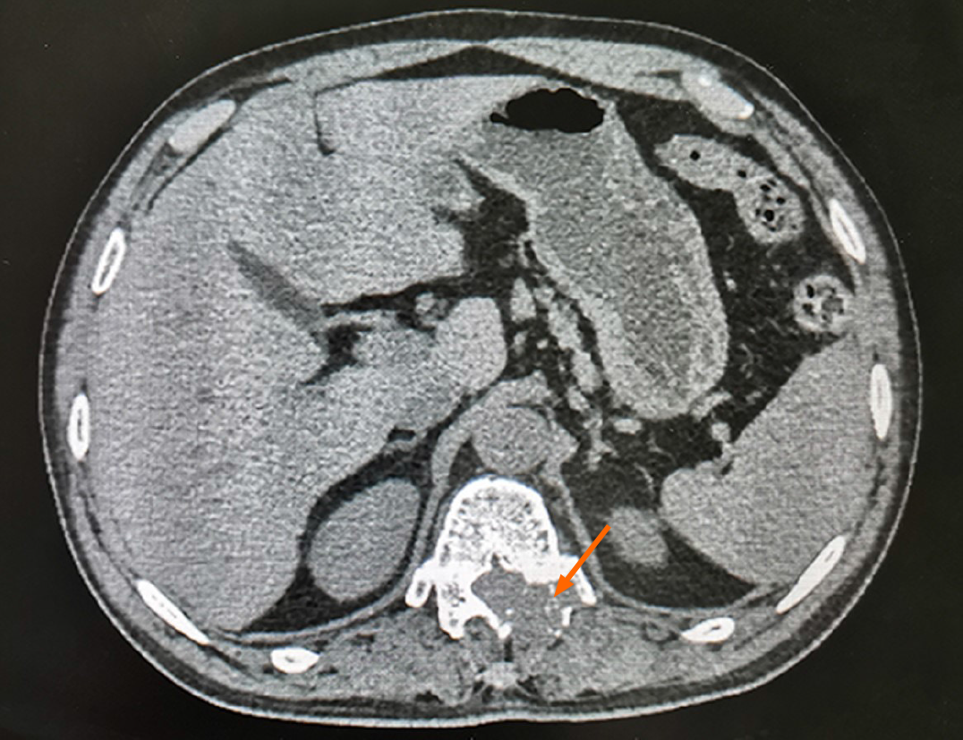
Figure 1 Preoperative computed tomography image revealing bone destruction of the left pedicle of the 12th thoracic vertebra.
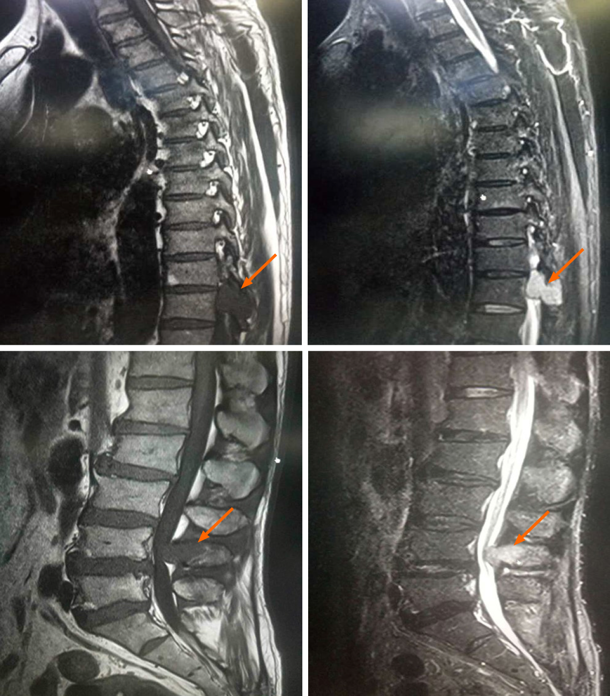
Figure 2 Preoperative sagittal magnetic resonance imaging revealing that T12 and L4 vertebral appendages showed obvious bone destruction.
A tumor in the spine compressed the spinal cord accompanied by spinal canal stenosis.
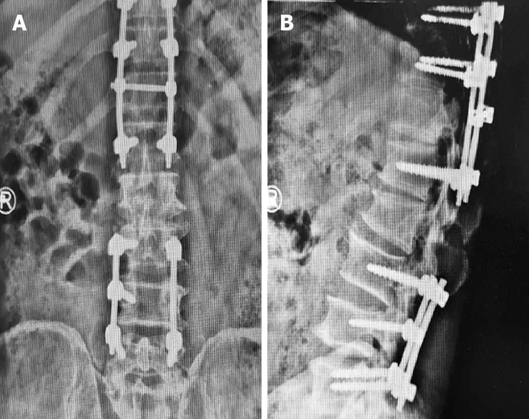
Figure 3 Frontal and lateral view of postoperative radiographs.
A: Frontal; B: Lateral.
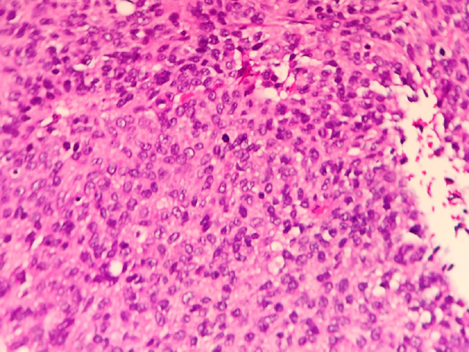
Figure 4 Postoperative biopsy revealing carcinomatous tissue.
- Citation: Kong Y, Ma XW, Zhang QQ, Zhao Y, Feng HL. Gastrointestinal stromal tumor with multisegmental spinal metastases as first presentation: A case report and review of the literature. World J Clin Cases 2021; 9(6): 1490-1498
- URL: https://www.wjgnet.com/2307-8960/full/v9/i6/1490.htm
- DOI: https://dx.doi.org/10.12998/wjcc.v9.i6.1490


