Copyright
©The Author(s) 2021.
World J Clin Cases. Feb 26, 2021; 9(6): 1408-1415
Published online Feb 26, 2021. doi: 10.12998/wjcc.v9.i6.1408
Published online Feb 26, 2021. doi: 10.12998/wjcc.v9.i6.1408
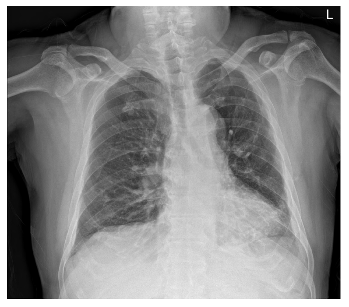
Figure 1 Preoperative chest radiograph.
Chest radiographs showed subsegmental atelectasis in the left lower lobe and mild cardiomegaly.
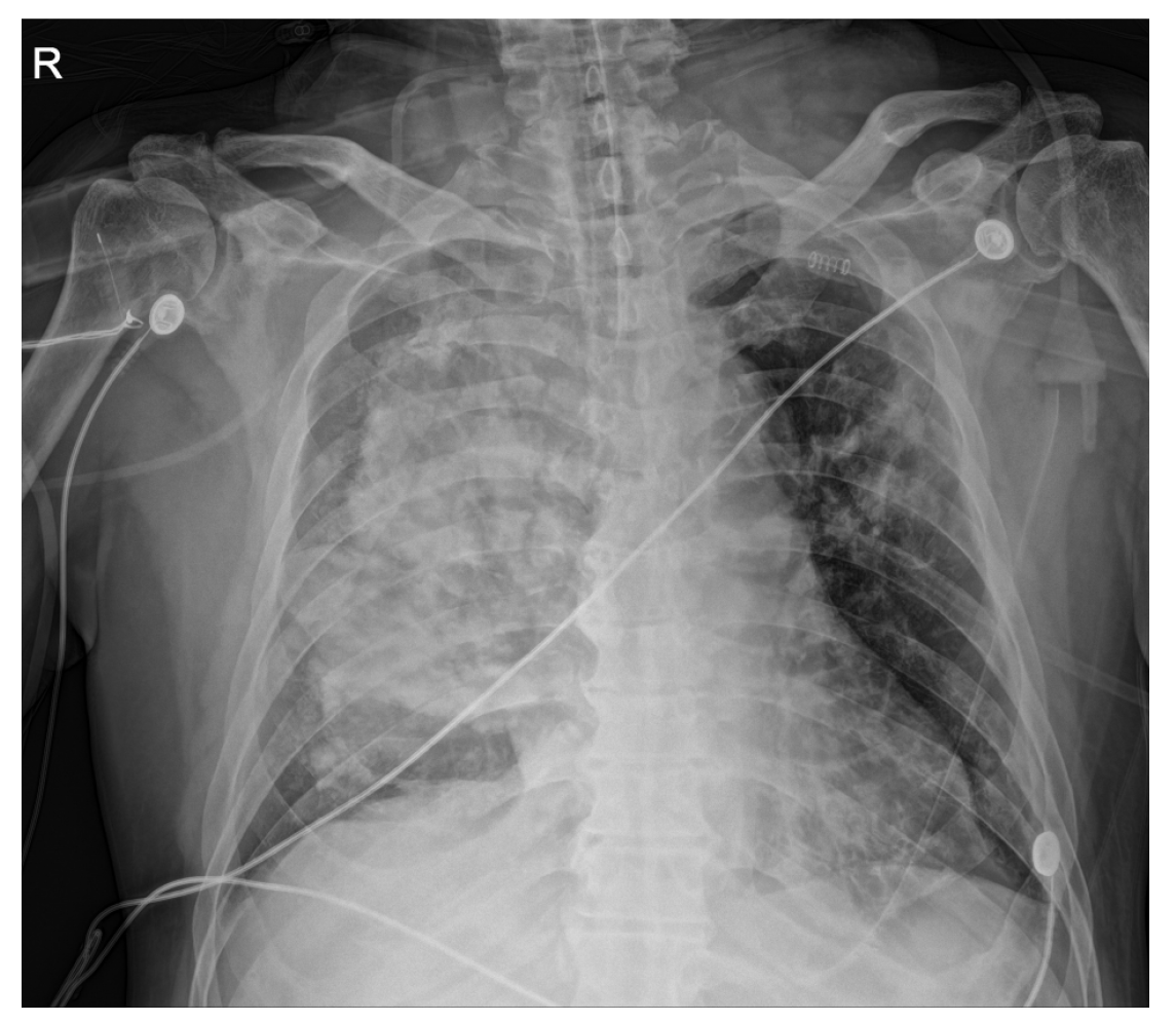
Figure 2 Postoperative chest radiograph at the intensive care unit.
Immediate postoperative chest X-ray revealed diffuse haziness of the entire right lung field.
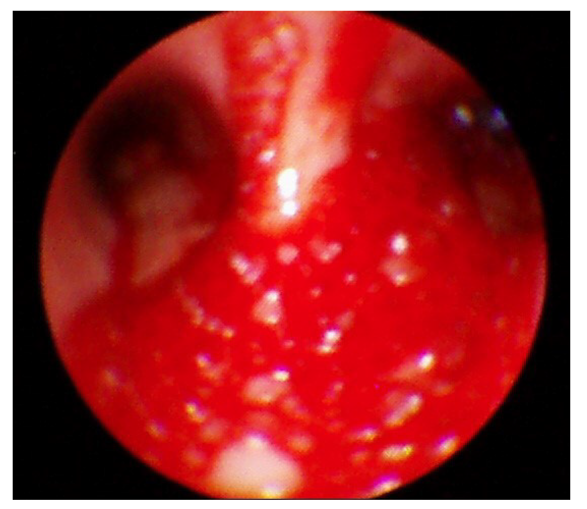
Figure 3 Postoperative bronchoscopic examination.
Postoperative bronchoscopy showed bloody secretions with red bubbles in the right lung.
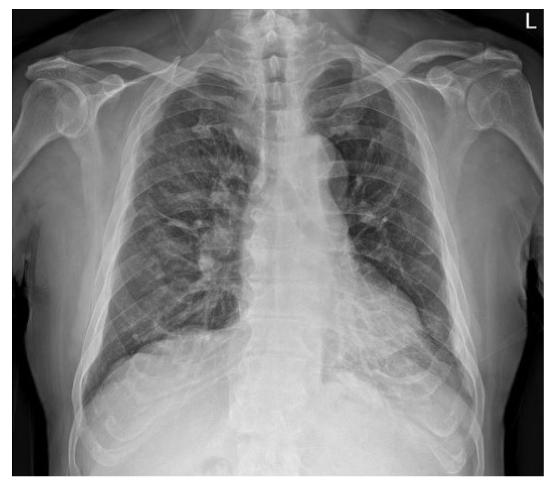
Figure 4 Postoperative chest radiographic examination on the eighth postoperative day.
After recovery, most of the radiologic haziness of the right lung had disappeared in the chest radiograph.
- Citation: Park HJ, Park SH, Woo UT, Cho SY, Jeon WJ, Shin WJ. Unilateral pulmonary hemorrhage caused by negative pressure pulmonary edema: A case report. World J Clin Cases 2021; 9(6): 1408-1415
- URL: https://www.wjgnet.com/2307-8960/full/v9/i6/1408.htm
- DOI: https://dx.doi.org/10.12998/wjcc.v9.i6.1408


