Copyright
©The Author(s) 2021.
World J Clin Cases. Feb 26, 2021; 9(6): 1402-1407
Published online Feb 26, 2021. doi: 10.12998/wjcc.v9.i6.1402
Published online Feb 26, 2021. doi: 10.12998/wjcc.v9.i6.1402
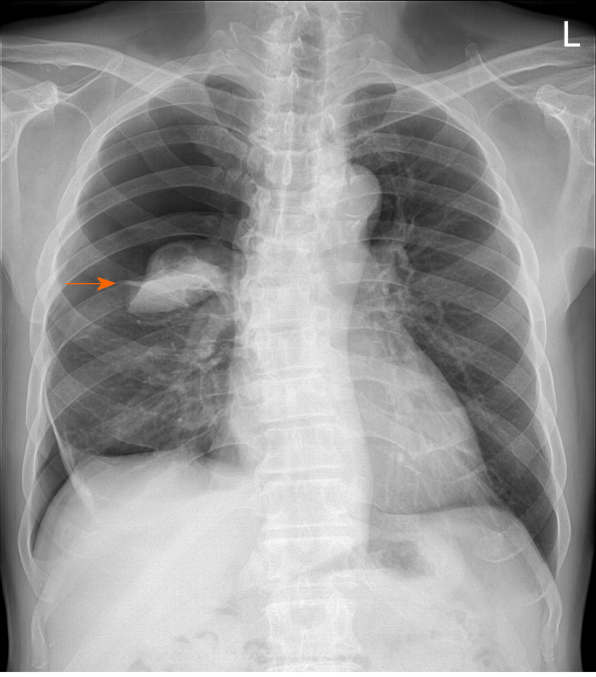
Figure 1 Visible pneumothorax (arrow) on an initial erect posteroanterior chest radiograph.
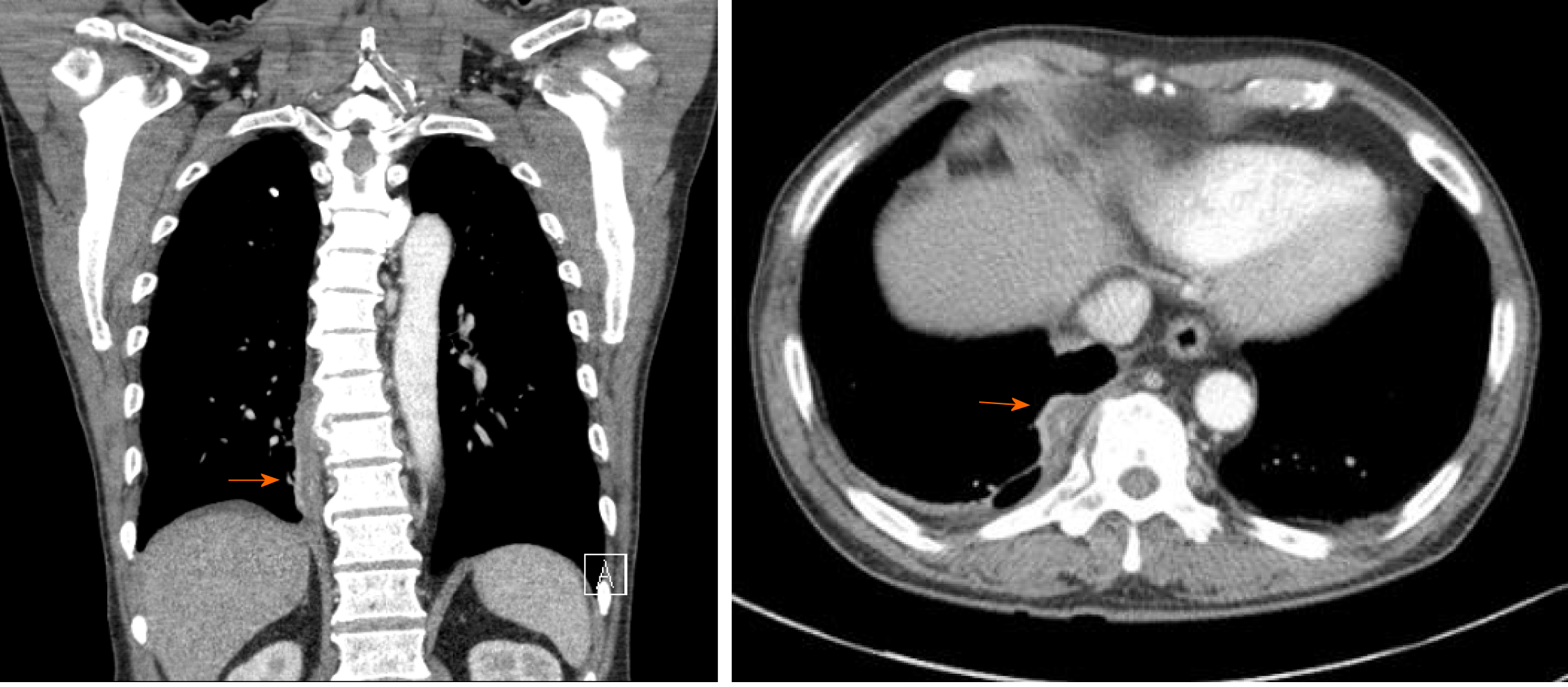
Figure 2 Initial chest computed tomography showing a paravertebral abscess (arrow) connected with the pleural effusion.
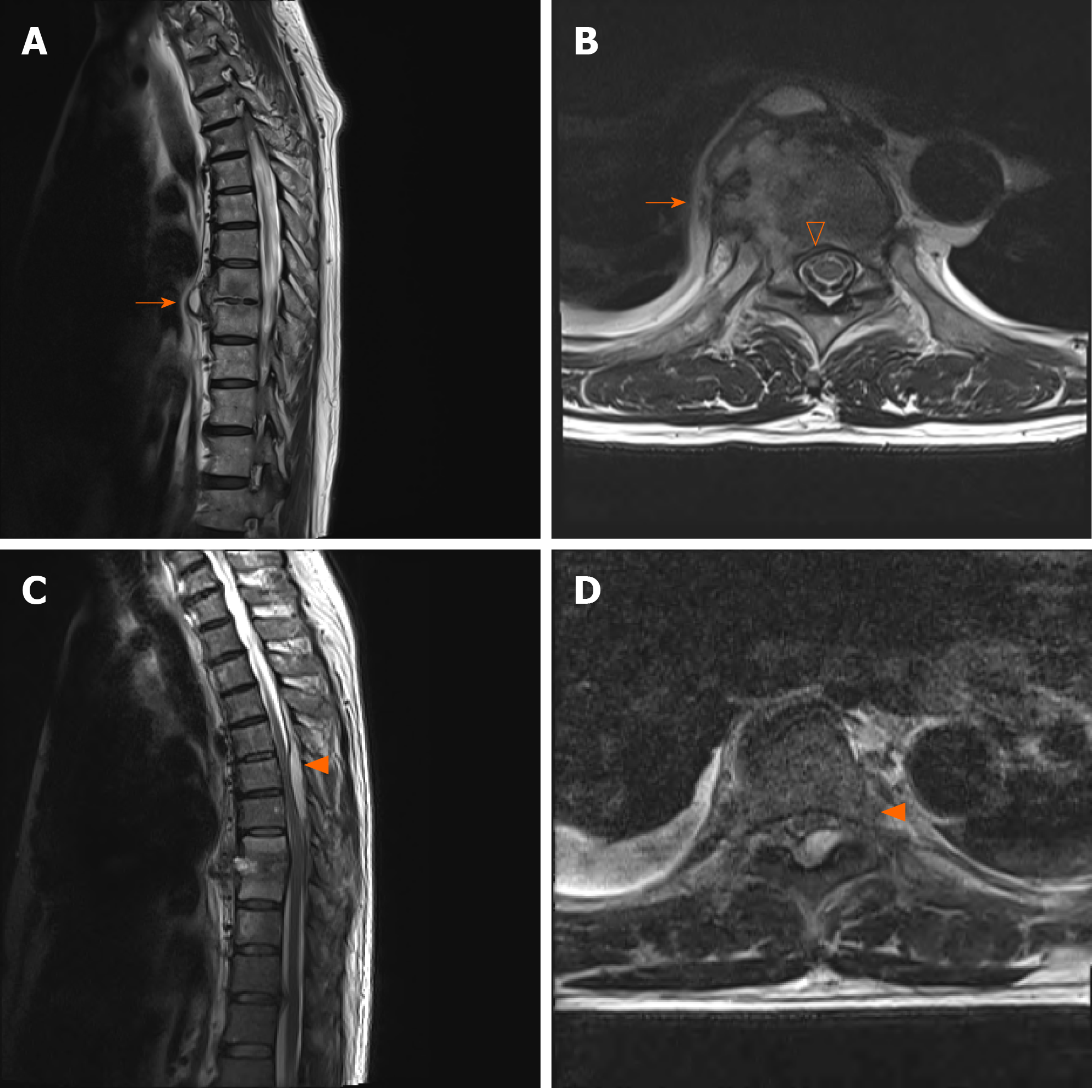
Figure 3 Magnetic resonance imaging of the thoracic spine.
A and B: Initial magnetic resonance imaging (MRI) scan showing a paravertebral abscess with extension to T8/9 disc, right rib (arrow), and anterior epidural space (empty arrowhead); C and D: Follow-up MRI scan showing an epidural abscess (arrowhead) at the T4-7 Level with spinal cord compression at T6-7.
- Citation: Cho MK, Lee BJ, Chang JH, Kim YM. Thoracic pyogenic infectious spondylitis presented as pneumothorax: A case report. World J Clin Cases 2021; 9(6): 1402-1407
- URL: https://www.wjgnet.com/2307-8960/full/v9/i6/1402.htm
- DOI: https://dx.doi.org/10.12998/wjcc.v9.i6.1402


