Copyright
©The Author(s) 2021.
World J Clin Cases. Feb 26, 2021; 9(6): 1379-1385
Published online Feb 26, 2021. doi: 10.12998/wjcc.v9.i6.1379
Published online Feb 26, 2021. doi: 10.12998/wjcc.v9.i6.1379
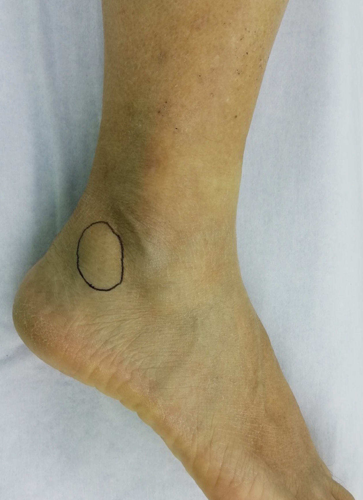
Figure 1 A mass (black circle) located at the posterior lower margin of the right lateral malleolus tip.
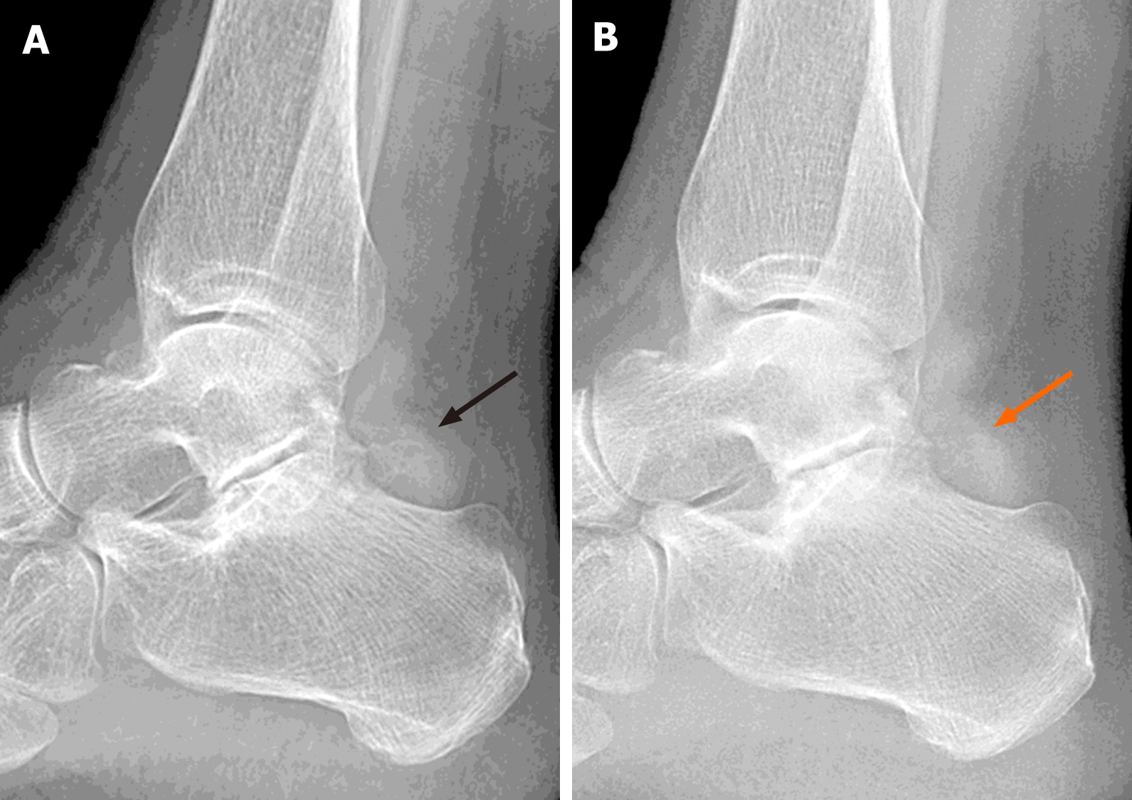
Figure 2 X-rays revealed osteophyte formation at the posterior margin of the subtalar joint and some irregular soft tissue calcification.
A and B: The irregular soft tissue calcification (A, black arrow) was more apparent than at 18 mo before the second visit (B, orange arrow).
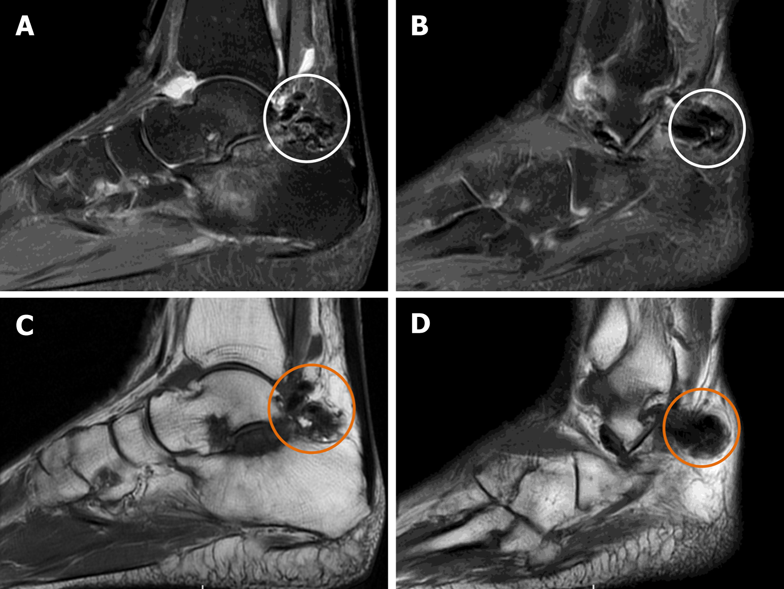
Figure 3 Magnetic resonance imaging.
A and B: Sagittal T2-weighted images showed a low-intensity mass (white circle), which protruded from the subtalar joint into the posterior soft tissue; C and D: Sagittal T1-weighted images showed a low-intensity mass (orange circle) in the corresponding area. Besides that, mild degeneration of cartilage was observed in the subtalar joint, without apparent bony erosion.
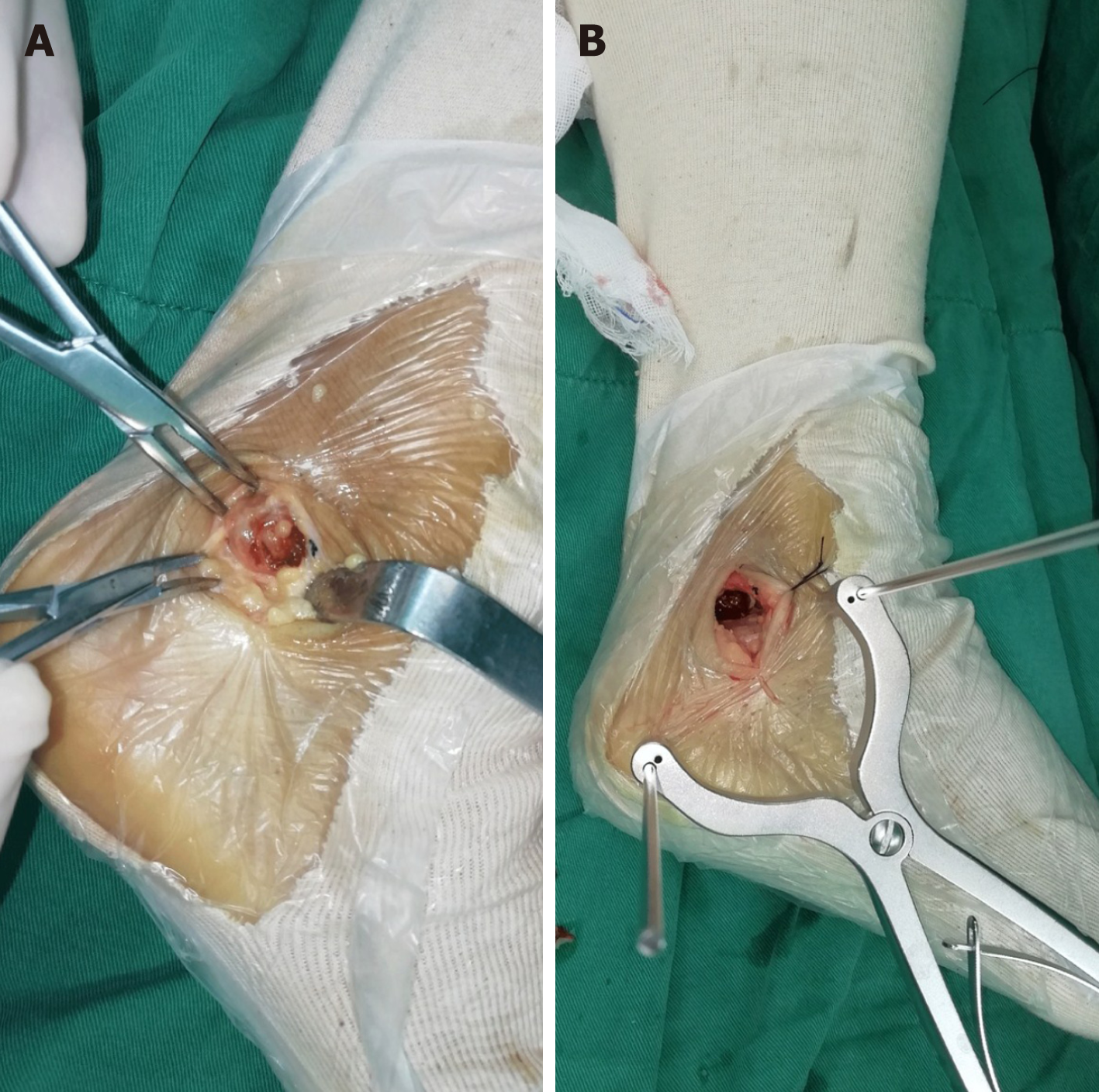
Figure 4 Surgical images.
A: Deep brown soft tissue was found during the operation; B: We gradually retracted the Steinmann pin until sufficient vision of the subtalar joint was achieved, and some of the cartilage was found to have mild degeneration.
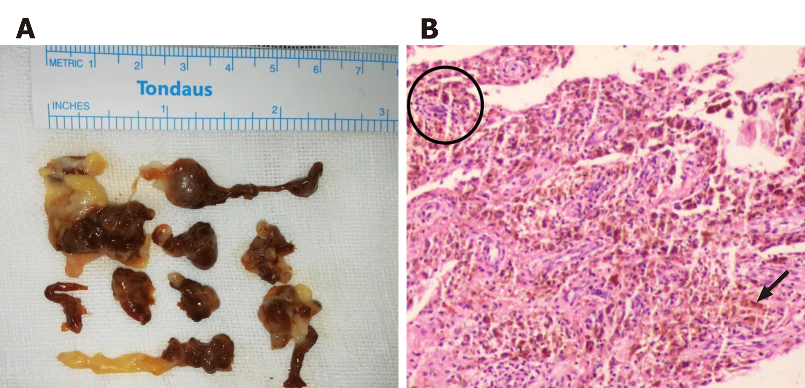
Figure 5 The postoperative histological examination images.
A: Dark brown and yellow soft pathological tissue mixed with a small amount of cartilaginous tissue; B: The pathological synovial tissue presented as villous nodular hyperplasia, with hemosiderin deposition (black arrow). Numerous multinucleated giant cells stained with hemosiderin (black circle).
- Citation: Zhao WQ, Zhao B, Li WS, Assan I. Subtalar joint pigmented villonodular synovitis misdiagnosed at the first visit: A case report. World J Clin Cases 2021; 9(6): 1379-1385
- URL: https://www.wjgnet.com/2307-8960/full/v9/i6/1379.htm
- DOI: https://dx.doi.org/10.12998/wjcc.v9.i6.1379


