Copyright
©The Author(s) 2021.
World J Clin Cases. Feb 26, 2021; 9(6): 1343-1352
Published online Feb 26, 2021. doi: 10.12998/wjcc.v9.i6.1343
Published online Feb 26, 2021. doi: 10.12998/wjcc.v9.i6.1343
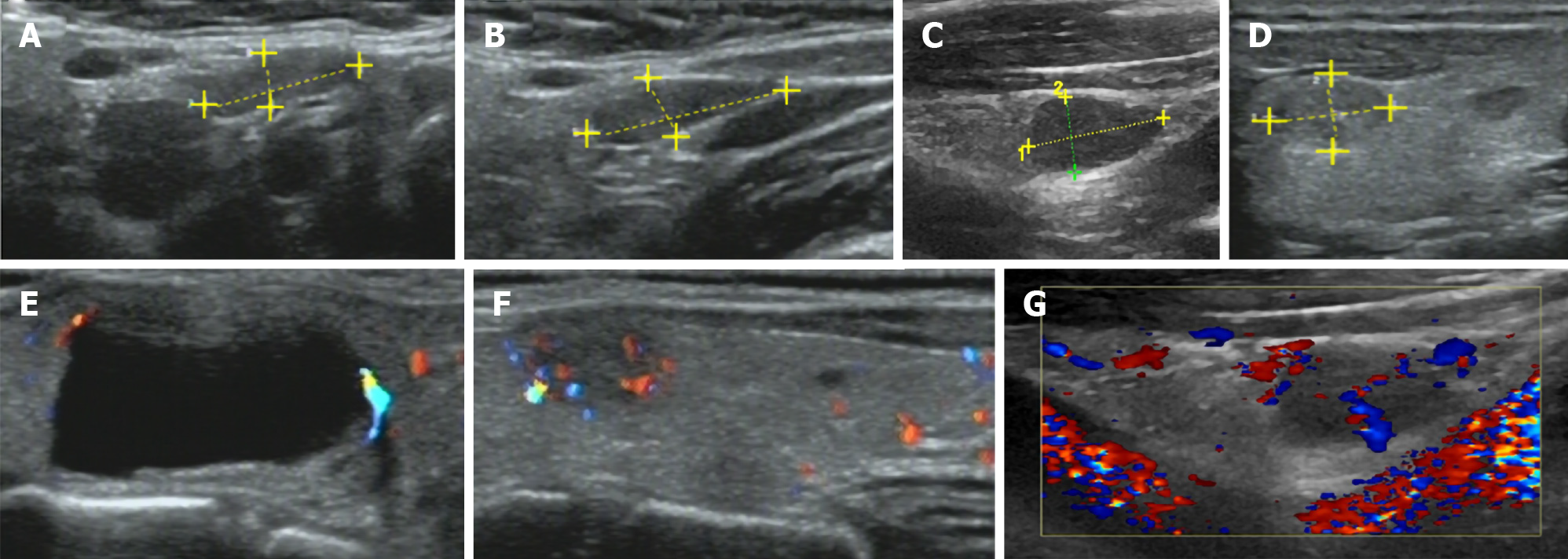
Figure 1 Ultrasound images.
A-D: Ultrasound (US) suggested multiple hypoechoic nodules in bilateral lobes of the thyroid, with clear boundaries; E-G: US indicated the dotted blood flow signal in nodules.
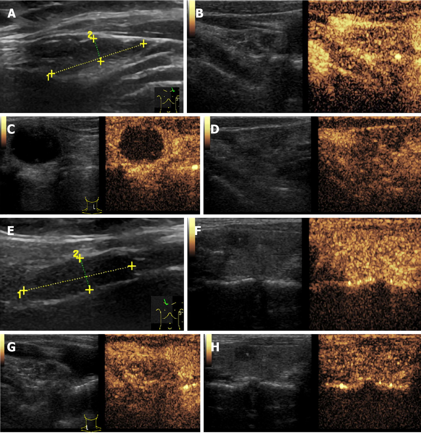
Figure 2 Ultrasound images.
A/E: Ultrasound suggested enlargement of cervical lymph nodes; B-D/F-H: Further contrast-enhanced ultrasound suggested uneven enhancement of enlarged lymph nodes from the medulla to cortex, with slightly lower medulla enhancement, suggesting that enlarged lymph nodes were mostly reactive hyperplasia.
Figure 3 Fine needle aspiration cytology.
A and B: Right thyroid: There were many follicular cell masses, arranged in branching or thick papillary shape, the nuclei were large and pale, and the inclusion bodies were visible. These findings suggested that a papillary thyroid carcinoma [the Bethesda system for reporting thyroid cytopathology (TBSRTC) V] should be suspected; C: Left thyroid: A small number of cell clusters, cell arrangement disorder, large nucleus, obvious pleomorphism, and chromatin fine structure were noted. These findings suggested that a malignancy (TBSRTC V, type indeterminate) should be suspected.
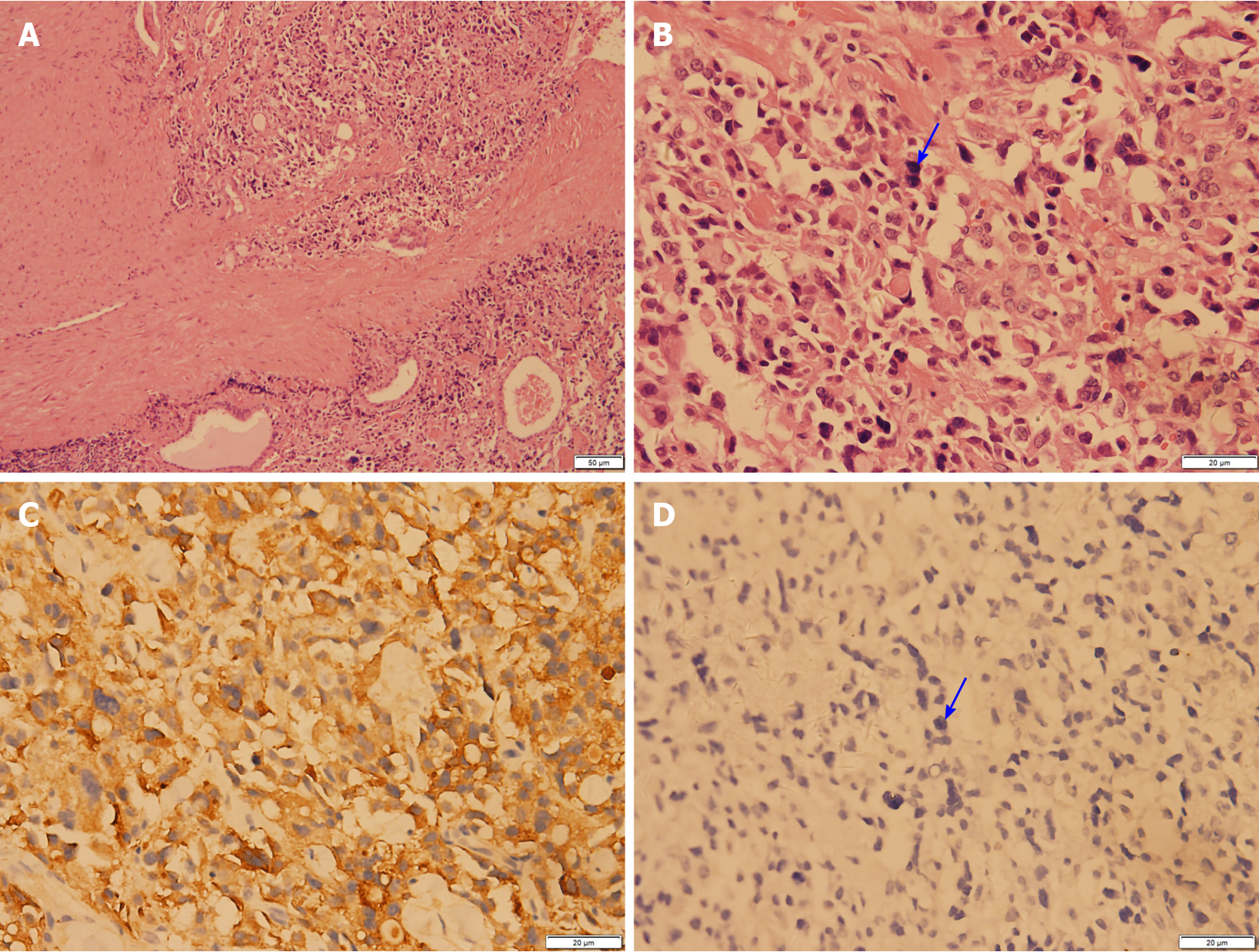
Figure 4 Paraffin pathology (right thyroid).
A and B: Photomicrographs showing hematoxylin-eosin (H&E) staining of a left thyroid nodule. The medullary thyroid carcinoma (MTC) component was mainly composed of irregular and solid nests of pleomorphic cells surrounded by a fibrovascular stroma with abundant amounts of acidophilic homogenous material, with large and polygonal cells, prominent nucleoli, and finely granular cytoplasm; C and D: The tumor cells of the MTC showed strong immunoreactivity for calcitonin and were negative for thyroglobulin.
Figure 5 Paraffin pathology (right thyroid).
A-C: Photomicrographs showing hematoxylin-eosin (H&E) staining of a right thyroid nodule.
- Citation: Gan FJ, Zhou T, Wu S, Xu MX, Sun SH. Do medullary thyroid carcinoma patients with high calcitonin require bilateral neck lymph node clearance? A case report. World J Clin Cases 2021; 9(6): 1343-1352
- URL: https://www.wjgnet.com/2307-8960/full/v9/i6/1343.htm
- DOI: https://dx.doi.org/10.12998/wjcc.v9.i6.1343


