Copyright
©The Author(s) 2021.
World J Clin Cases. Feb 16, 2021; 9(5): 1237-1246
Published online Feb 16, 2021. doi: 10.12998/wjcc.v9.i5.1237
Published online Feb 16, 2021. doi: 10.12998/wjcc.v9.i5.1237
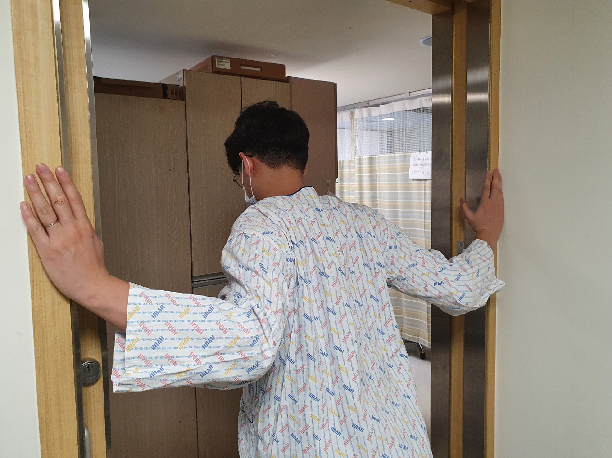
Figure 1 Stretching exercise of the pectoralis minor muscle.
Both shoulder joints were mildly extended, 90° externally rotated, and 45° abducted. Both scapulae were retracted and the elbow joints were bent approximately 90°. The patient placed his elbows on the wall right next to the door and pushed his trunk forward.
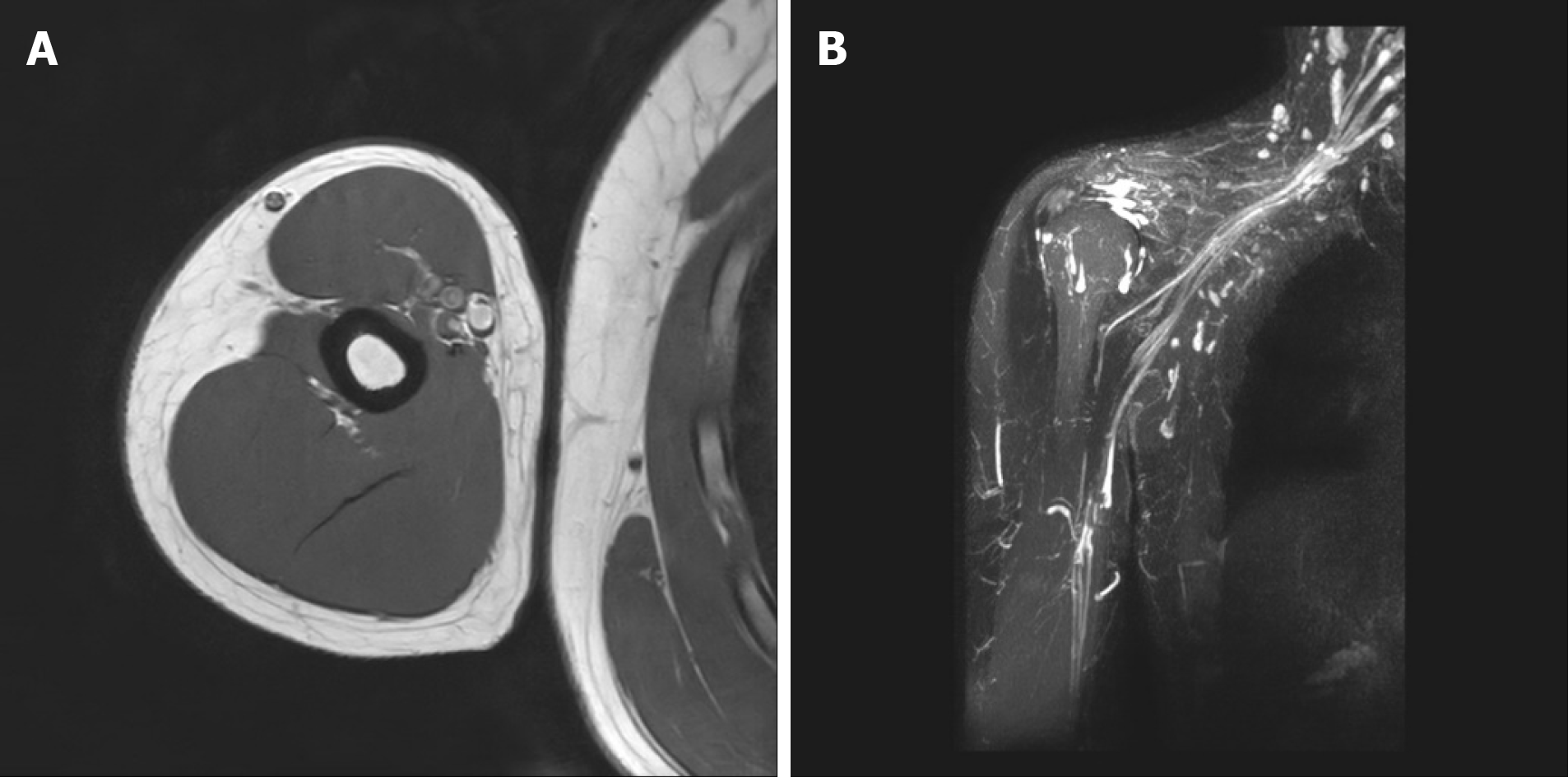
Figure 2 Right arm and brachial plexus magnetic resonance imaging.
A: Transverse view; B: Coronal view. Significant abnormality was not observed in the right brachial plexus and distal peripheral nerves.
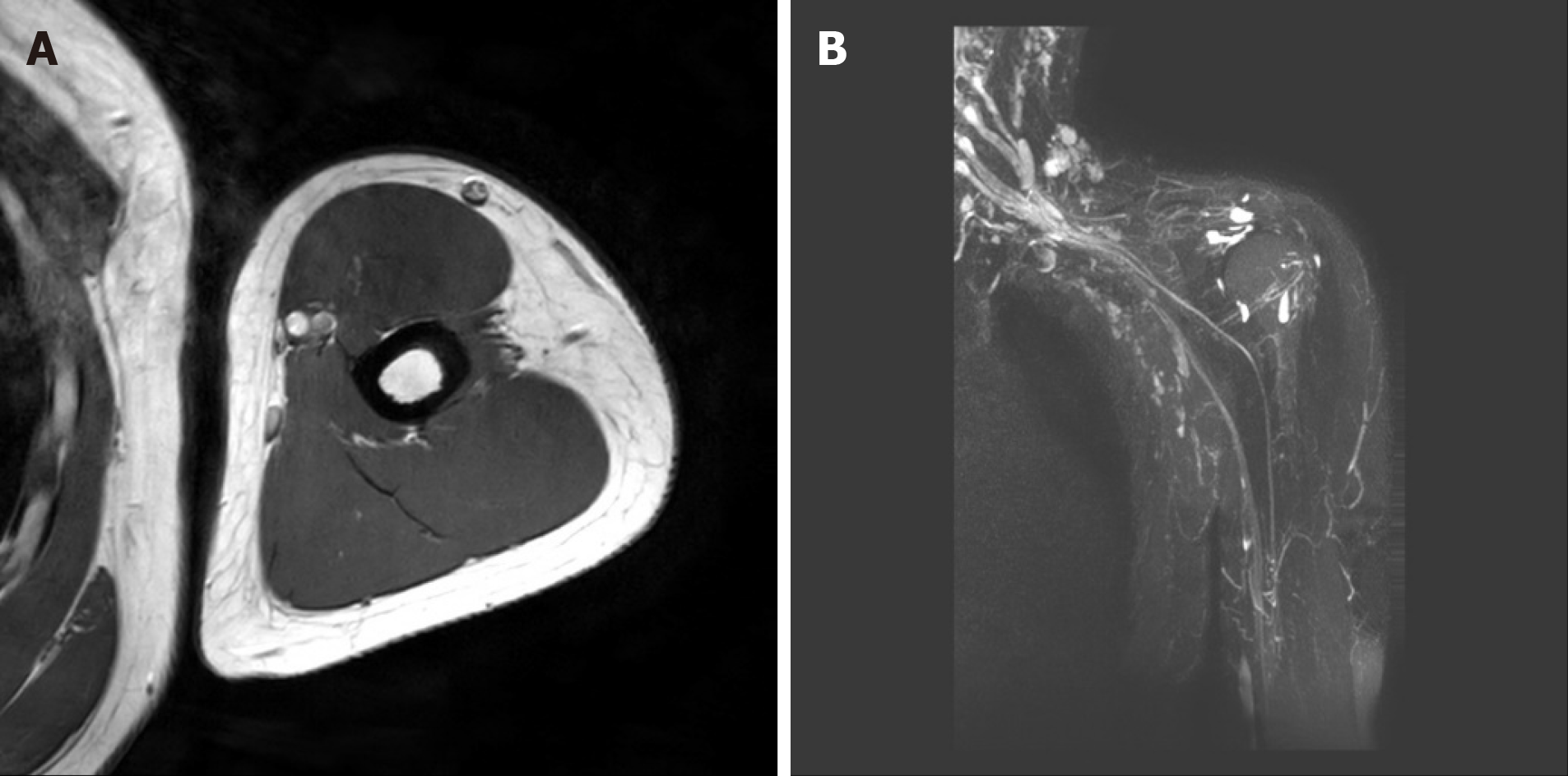
Figure 3 Left arm and brachial plexus magnetic resonance imaging.
A: Transverse view; B: Coronal view. Significant abnormality was not observed in the left brachial plexus and distal peripheral nerves.
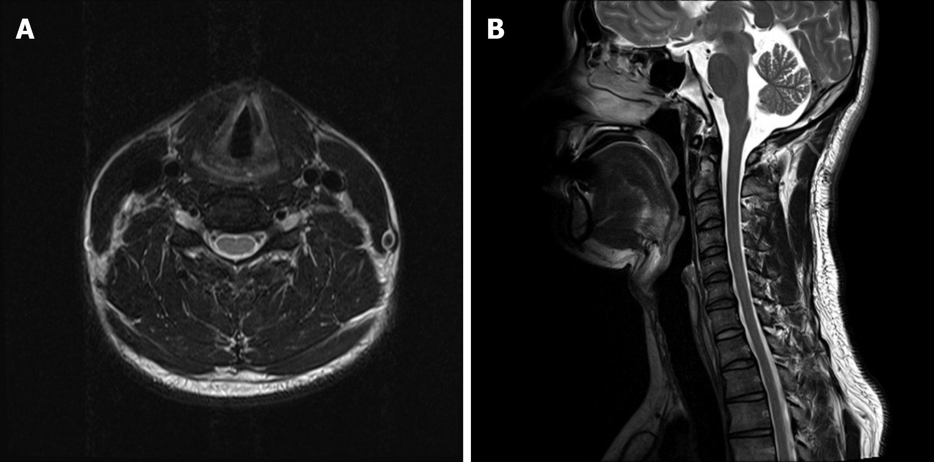
Figure 4 Cervical spine magnetic resonance imaging taken at a previous hospital.
A: Transverse view; B: Sagittal view. Specific abnormalities were not observed on cervical spine magnetic resonance imaging.
- Citation: Jung JW, Park YC, Lee JY, Park JH, Jang SH. Bilateral musculocutaneous neuropathy: A case report. World J Clin Cases 2021; 9(5): 1237-1246
- URL: https://www.wjgnet.com/2307-8960/full/v9/i5/1237.htm
- DOI: https://dx.doi.org/10.12998/wjcc.v9.i5.1237


