Copyright
©The Author(s) 2021.
World J Clin Cases. Feb 16, 2021; 9(5): 1228-1236
Published online Feb 16, 2021. doi: 10.12998/wjcc.v9.i5.1228
Published online Feb 16, 2021. doi: 10.12998/wjcc.v9.i5.1228
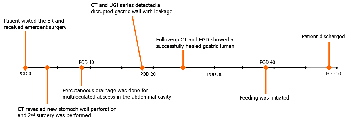
Figure 1 Timeline of care.
ER: Emergency room; CT: Computed tomography; UGI: Upper gastrointestinal; EGD: Esophagogastroduodenoscopy; POD: Postoperative day.
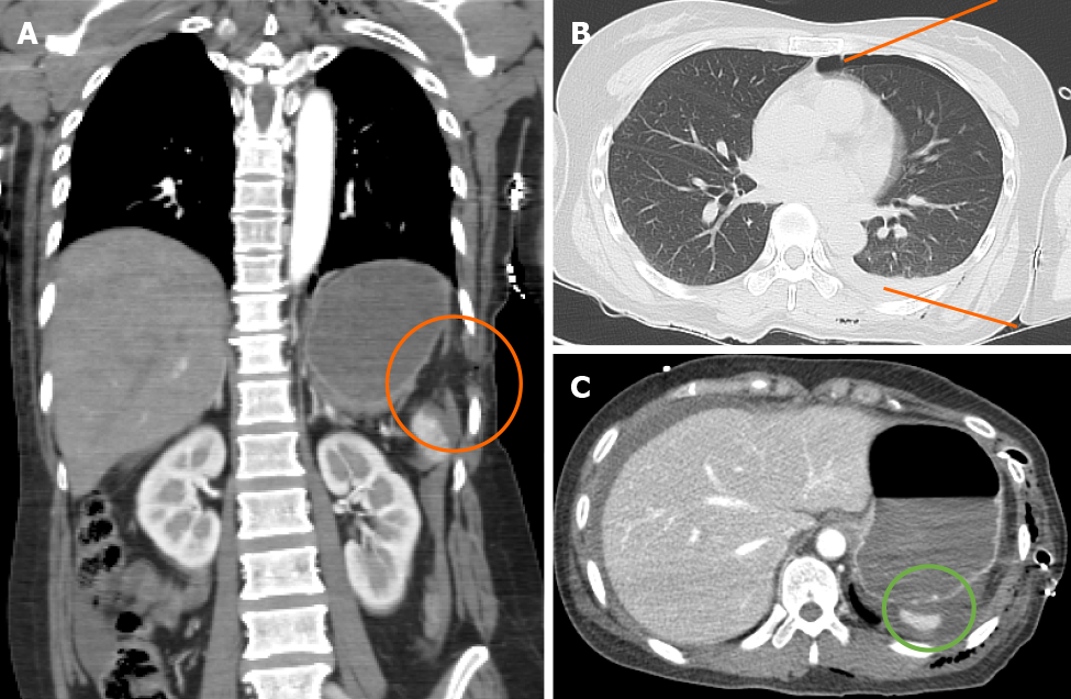
Figure 2 Initial computed tomography scan of the abdominal cavity.
A-C: This scan shows left hemidiaphragmatic injury with herniation (A, orange circle), left hemopneumothorax (B, orange line), and perisplenic hemorrhage without signs of gastric perforation (C, green circle).
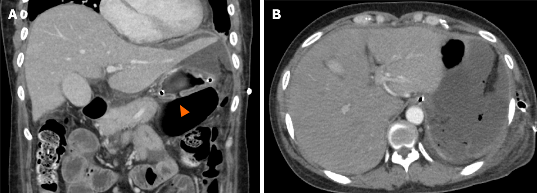
Figure 3 Follow-up computed tomography scan on 3rd postoperative day.
A and B: Free perforation of the upper stomach (A, orange arrowhead) with a hemoperitoneum and pneumoperitoneum (B) is noted.
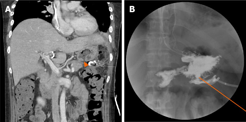
Figure 4 Radiologic findings of re-perforated gastric wall.
A and B: Computed tomography findings showing gastric wall re-perforation (A, orange arrowhead) and upper gastrointestinal series showing leakage of the gastric contents (B, orange line).
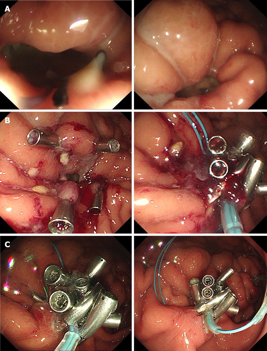
Figure 5 Primary closure of the perforated gastric wall with an endoscopic procedure.
A and B: Endoscopic findings of the re-perforated gastric lumen (A) and its primary closure with endoloops and clips using the modified purse-string technique (B); C: Final endoscopic findings on 25th postoperative day showing well-healed gastric wall.
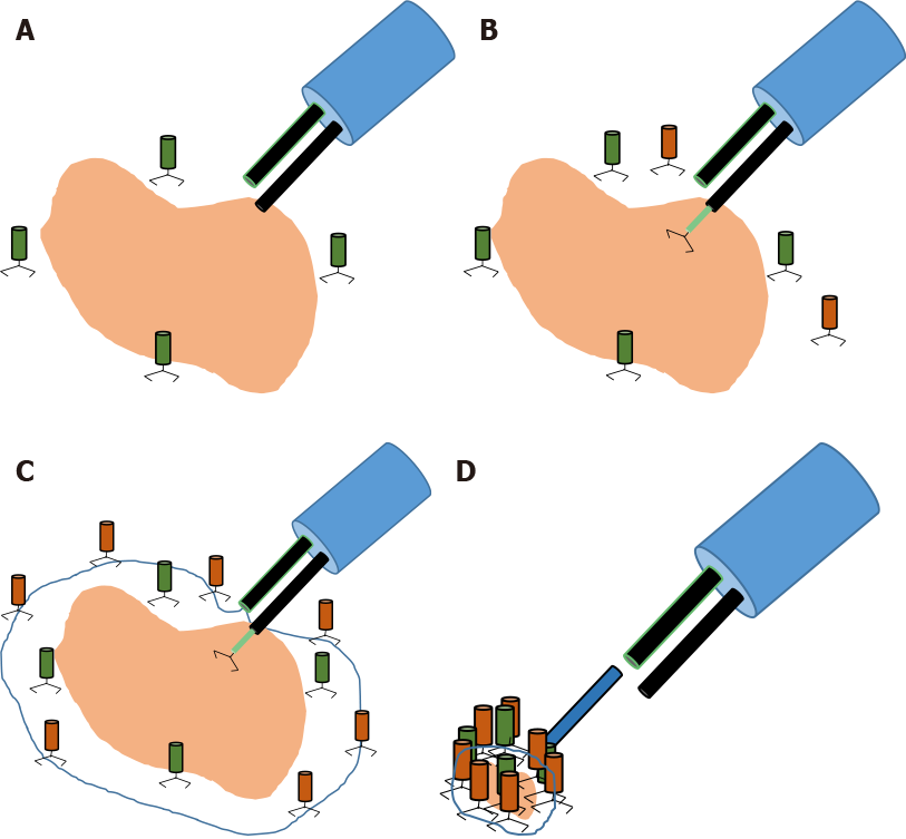
Figure 6 Schematic diagram of the purse-string technique with pillar clips.
A: After detection of the perforated stomach lumen and swollen edematous mucosa, pillar clips are fixed near the margin of the perforated wall; B and C: The endoloop is then placed at the outer margin of the pillar clips and is fixed with additional clips; D: Finally, the endoloop is fastened with the purse-string technique.
- Citation: Yoon JH, Jun CH, Han JP, Yeom JW, Kang SK, Kook HY, Choi SK. Endoscopic repair of delayed stomach perforation caused by penetrating trauma: A case report. World J Clin Cases 2021; 9(5): 1228-1236
- URL: https://www.wjgnet.com/2307-8960/full/v9/i5/1228.htm
- DOI: https://dx.doi.org/10.12998/wjcc.v9.i5.1228


