Copyright
©The Author(s) 2021.
World J Clin Cases. Feb 16, 2021; 9(5): 1221-1227
Published online Feb 16, 2021. doi: 10.12998/wjcc.v9.i5.1221
Published online Feb 16, 2021. doi: 10.12998/wjcc.v9.i5.1221
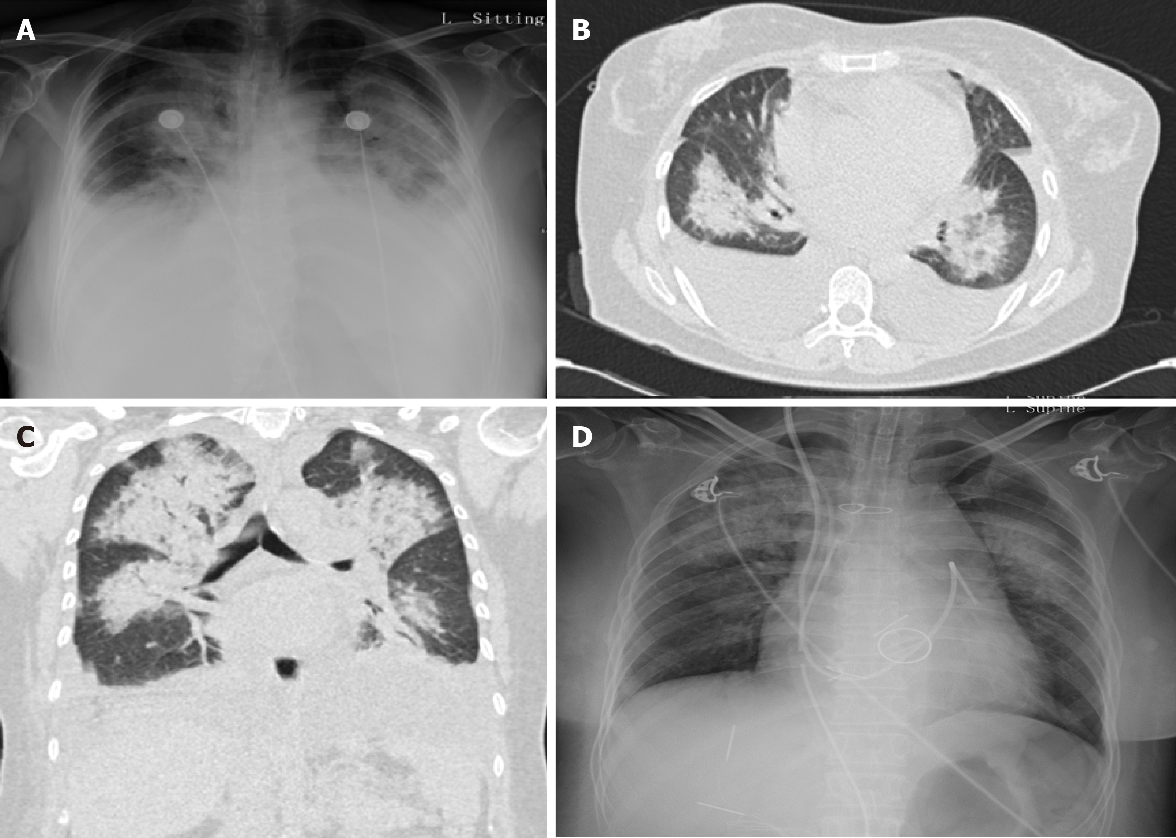
Figure 1 Chest X-ray and computed tomography images.
A: Chest X-ray upon presentation showed diffuse pulmonary edema and bilateral pleural effusion; B and C: Extensive pulmonary edema and bilateral pleural effusion were also observed on computed tomography scanning; D: Chest X-ray showed significant amelioration of pulmonary edema and pleural effusion following replacement of the infected valve.
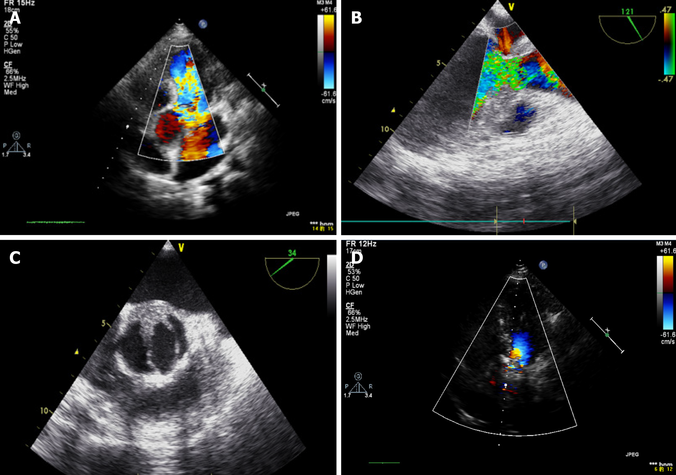
Figure 2 Transthoracic and transesophageal echocardiography images.
A: Immediate transthoracic echocardiography revealed severe aortic regurgitation without left ventricular enlargement; B: Transesophageal echocardiography also revealed severe aortic regurgitation; C: Vegetations attached to the bicuspid aortic valve were clearly visible; D: Postoperative transthoracic echocardiography revealed that regurgitation of the aortic prosthetic valve was significantly reduced.
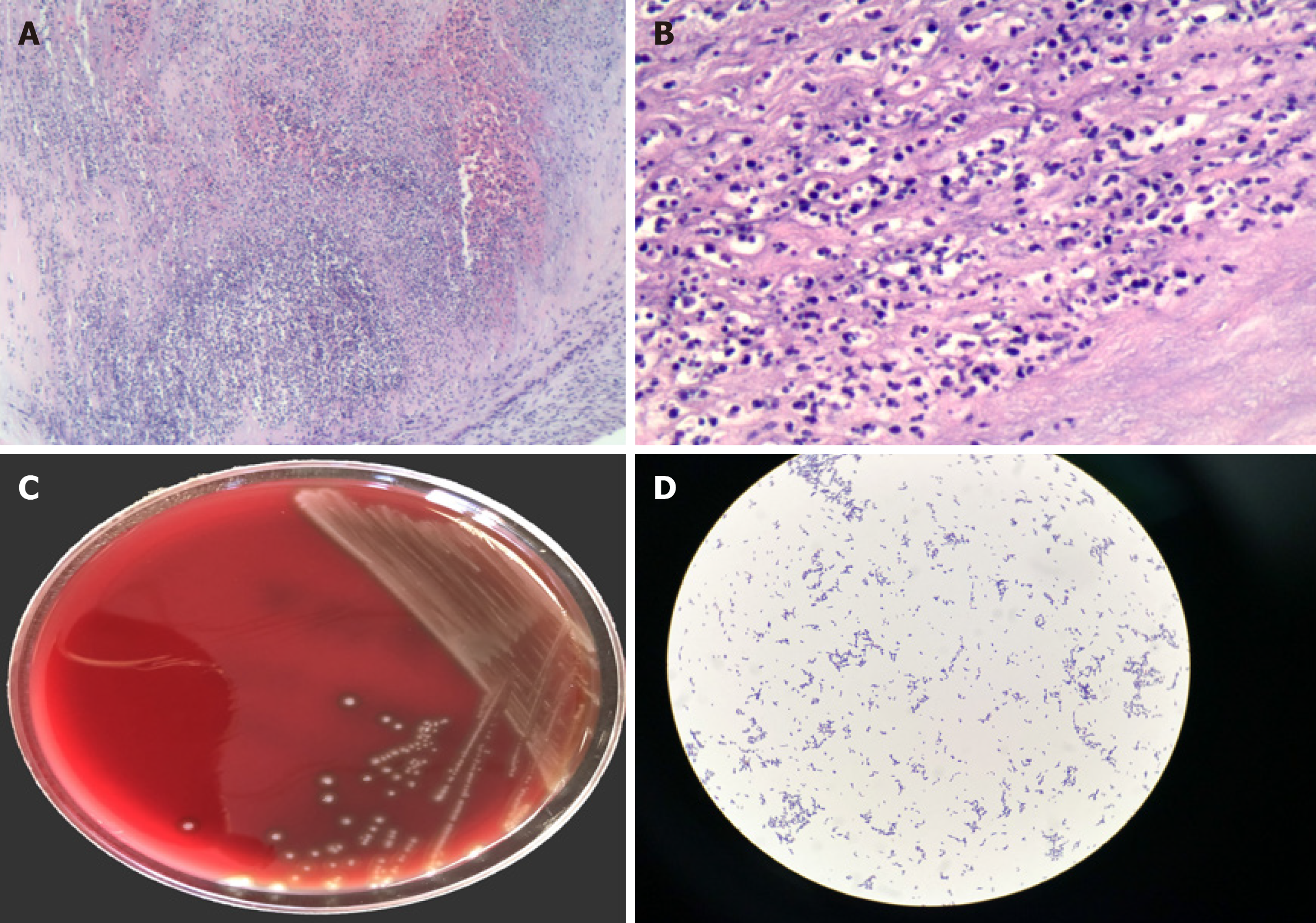
Figure 3 Histopathology of valvular specimens and micro-organism culture of excised tissues.
A: Histopathological examination demonstrated inflammatory infiltrates containing large numbers of neutrophils (Hematoxylin & eosin staining, × 40); B: Active inflammation with abundant neutrophilic infiltration could also be seen at high power view (Hematoxylin & eosin staining, × 200); C: Positive culture of Streptococcus sanguinis isolated from the resected samples; D: Microscopic appearance of cultured Streptococcus sanguinis.
- Citation: Hou C, Wang WC, Chen H, Zhang YY, Wang WM. Infective bicuspid aortic valve endocarditis causing acute severe regurgitation and heart failure: A case report. World J Clin Cases 2021; 9(5): 1221-1227
- URL: https://www.wjgnet.com/2307-8960/full/v9/i5/1221.htm
- DOI: https://dx.doi.org/10.12998/wjcc.v9.i5.1221


