Copyright
©The Author(s) 2021.
World J Clin Cases. Feb 16, 2021; 9(5): 1204-1209
Published online Feb 16, 2021. doi: 10.12998/wjcc.v9.i5.1204
Published online Feb 16, 2021. doi: 10.12998/wjcc.v9.i5.1204
Figure 1 Under general anesthesia, an incision was made via a parallel path from the back of the right ear to the submandibular area to explore and to protect the facial nerve, and the tumors in the deep surface of the parotid gland and the right parapharyngeal space were completely removed.
A: A superficial tumor that compressed the parotid gland; B: A deep tumor in the parapharyngeal space; C: Resected tumors compared with a 10 mL syringe.
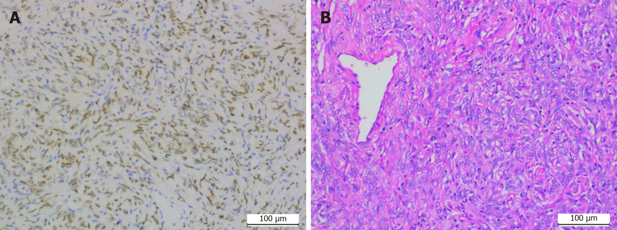
Figure 2 Immunohistochemistry of signal transducer and activator of transcription 6 (Elivision × 100) (A) and hematoxylin and eosin staining of tumor cells (× 100) (B).
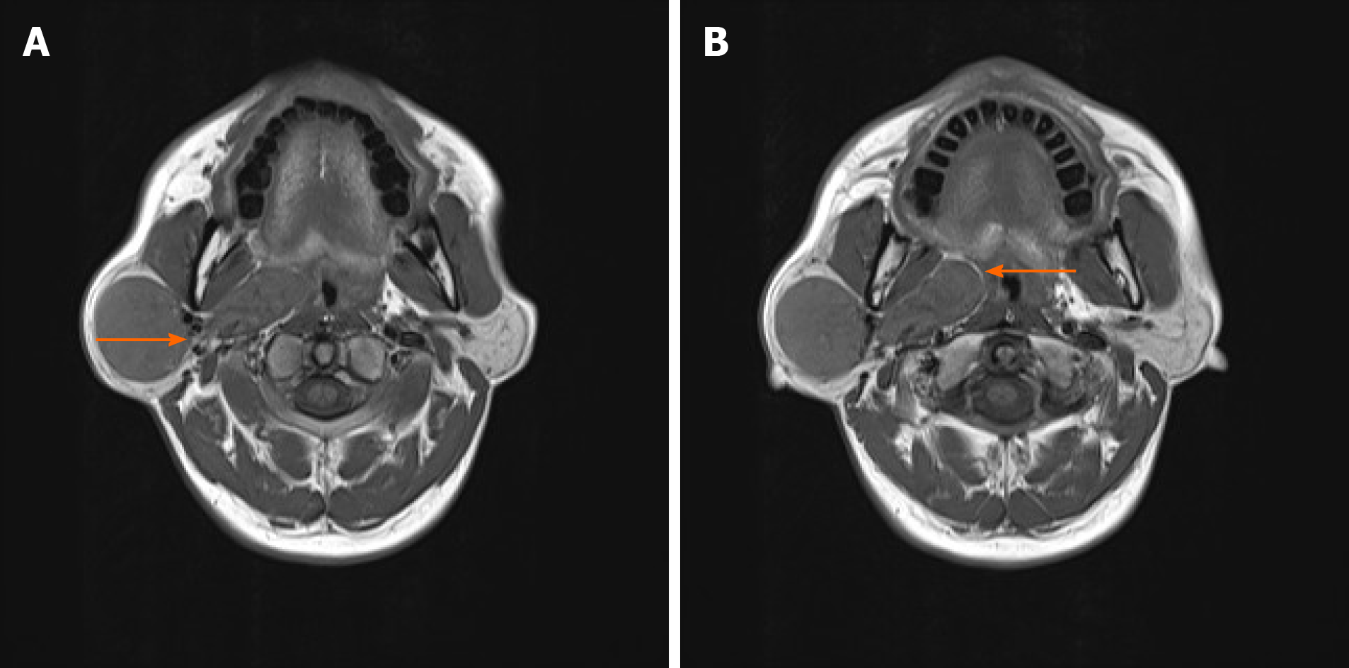
Figure 3 A clear boundary between the parotid gland tumor and the parotid gland is observed, and the two tumors are connected at the point indicated by the arrow (A) and fatty inclusion sign of a schwannoma is indicated by the arrow (B).
- Citation: Li YN, Li CL, Liu ZH. Dumbbell-shaped solitary fibrous tumor in the parapharyngeal space: A case report. World J Clin Cases 2021; 9(5): 1204-1209
- URL: https://www.wjgnet.com/2307-8960/full/v9/i5/1204.htm
- DOI: https://dx.doi.org/10.12998/wjcc.v9.i5.1204


