Copyright
©The Author(s) 2021.
World J Clin Cases. Feb 16, 2021; 9(5): 1196-1203
Published online Feb 16, 2021. doi: 10.12998/wjcc.v9.i5.1196
Published online Feb 16, 2021. doi: 10.12998/wjcc.v9.i5.1196
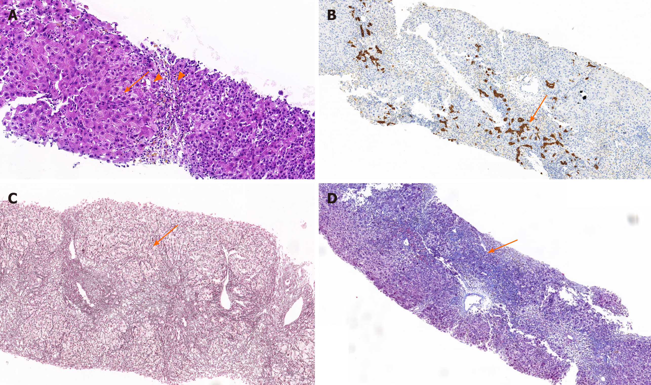
Figure 1 Imaging examinations results.
A: In the hematoxylin and eosin staining of liver tissue, the structure of hepatic lobules was unclear. The hematoxylin and eosin staining showed varying degrees of hepatocyte edema with moderate to severe piecemeal necrosis and scattered eosinophilic bodies (arrows). Small bile ducts in the portal area were accompanied by lymphocyte-predominant inflammation and a small amount of eosinophil infiltration (filled triangles); B: The proliferation of small bile ducts in the portal area were obvious (arrows) with more infiltration of lymphocytes and plasma cells and positive marker detection including hepatitis B surface antigen (+), HBcAg (+-), bile duct epithelium (CK19+), IgG (+), and IgG4 (-) combined hepatocellular and cholangiocarcinoma. Findings were consistent with mild chronic hepatitis compatible with hepatitis B virus infection, without histological stigmata of autoimmunity; C: Mesh fiber staining of liver tissue showed that the hepatic lobular stent was significantly damaged (arrows). The characteristic floral ring structure of autoimmune hepatitis was not seen; D: Masson staining of liver tissue showed the portal area was enlarged with fibrosis, the formation of the bridging fiber band was visible in the focal area, and collagen fiber was proliferating the portal area (arrows).
- Citation: Zhang X, Xie QX, Zhao DM. Negative conversion of autoantibody profile in chronic hepatitis B: A case report. World J Clin Cases 2021; 9(5): 1196-1203
- URL: https://www.wjgnet.com/2307-8960/full/v9/i5/1196.htm
- DOI: https://dx.doi.org/10.12998/wjcc.v9.i5.1196


