Copyright
©The Author(s) 2021.
World J Clin Cases. Feb 16, 2021; 9(5): 1168-1174
Published online Feb 16, 2021. doi: 10.12998/wjcc.v9.i5.1168
Published online Feb 16, 2021. doi: 10.12998/wjcc.v9.i5.1168
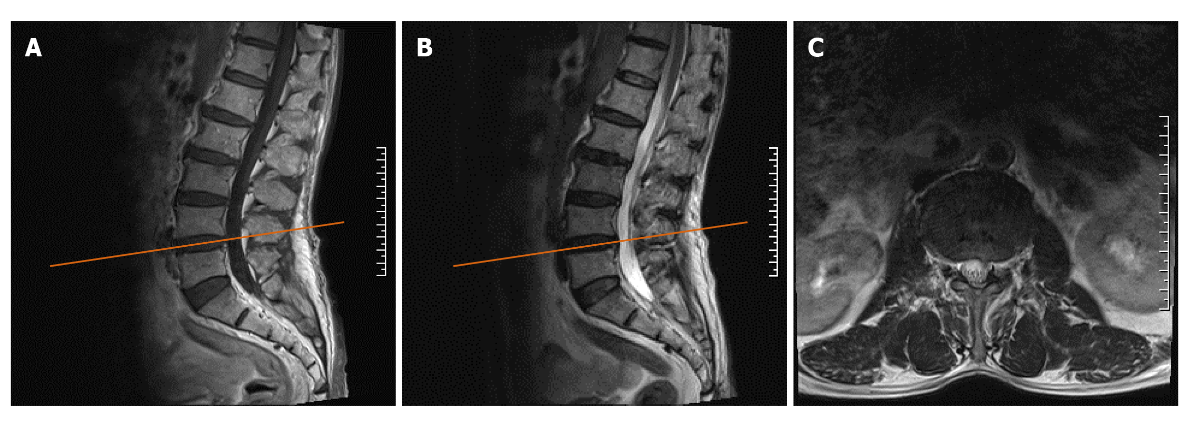
Figure 1 Plain magnetic resonance imaging of the lumbar before the operation.
A: T1 sagittal image showing intervertebral disc bulging at the L4/5 level, which caused slight compression of the anterior edge of the dural sac but not to the extent that causes neurological symptoms; B: T2 sagittal image of the L4/5 level; C: The horizontal image correlated with the L4/5 intervertebral space, as indicated by the yellow scout line in the panel.
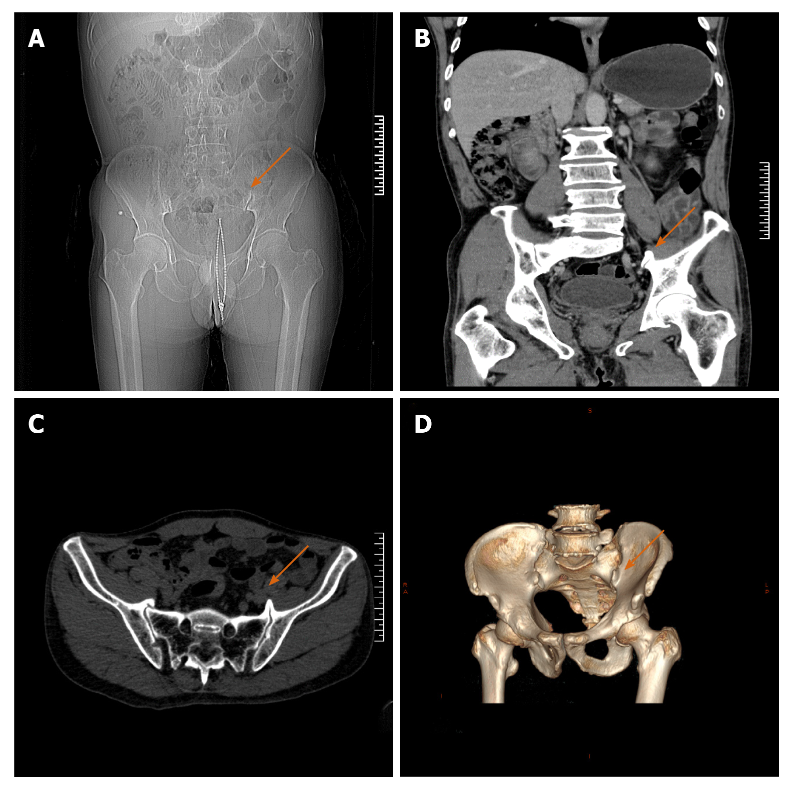
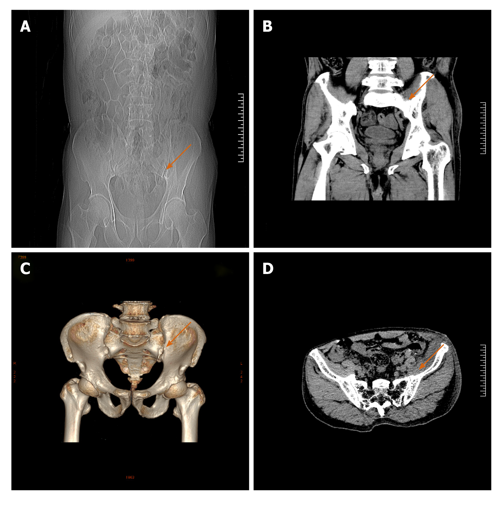
Figure 2 Pelvic computed tomography scan plus 3D reconstruction before the operation.
A: Anteroposterior radiograph of the pelvis; B: Coronal computed tomography scan showing the osteophyte in the left sacroiliac joint, as indicated by the orange arrow; C: Horizontal scan showing that the left osteophyte was more prominent; D: 3D reconstruction revealing the osteophytes that were in the anterior lower part of both sacroiliac joints, with the left osteophyte being larger, as indicated by the orange arrow in the figure.
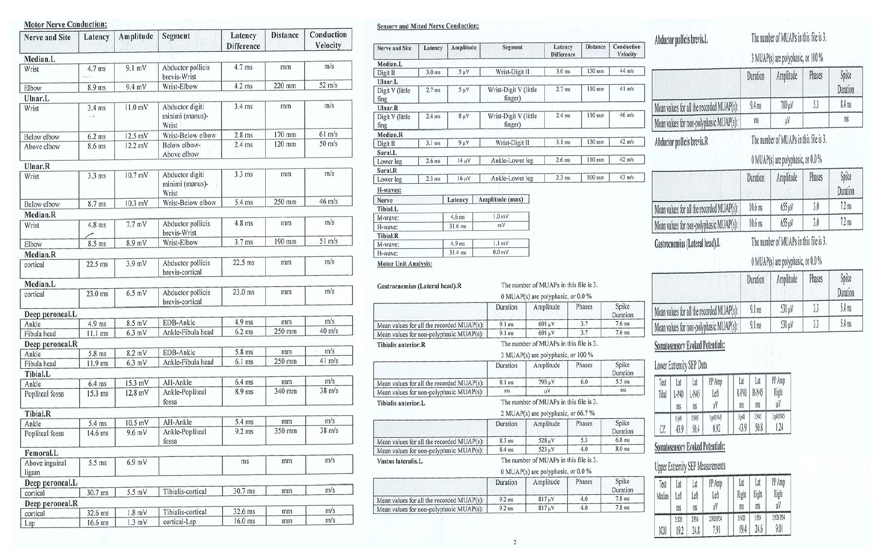
Figure 3 Electromyography and nerve conduction velocity of both lower extremities.
Electrophysiology showed that the P40 conduction latency time of lower extremities deep sensory pathway was normal while the amplitude was reduced and the waveform was poor, which means peripheral nerve injury.
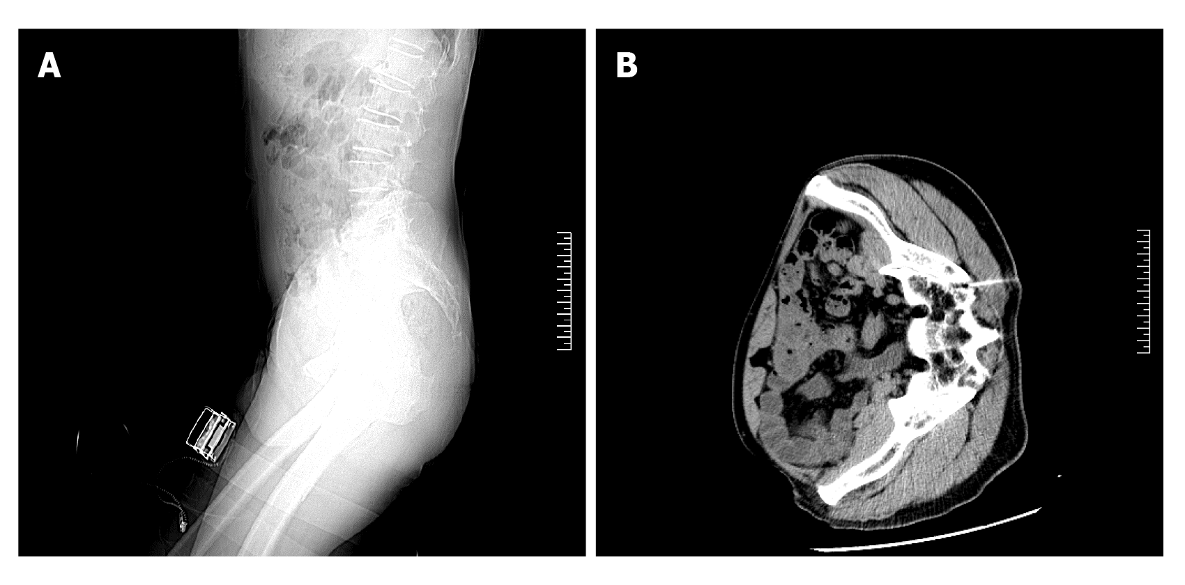
Figure 4 Computed tomography-guided diagnostic obturator nerve local block.
A: Sagittal image showing that computed tomography-guided posterior approach for obturator nerve block was demonstrating; B: Horizontal image showing the puncture needle was entering the sacroiliac joint space.
Figure 5 Pelvic computed tomography scan plus 3D reconstruction after the operation.
A: Anteroposterior radiograph of the pelvis; B: Coronal computed tomography scan showed the osteophyte has been removed; C: 3D reconstruction showed that the osteophyte from the anterior lower part of the left sacroiliac joint has been completely removed and the articular surface was flat while the osteophyte of the right was still existing, as indicated by the orange arrow; D: Horizontal scan of the pelvic after surgery.
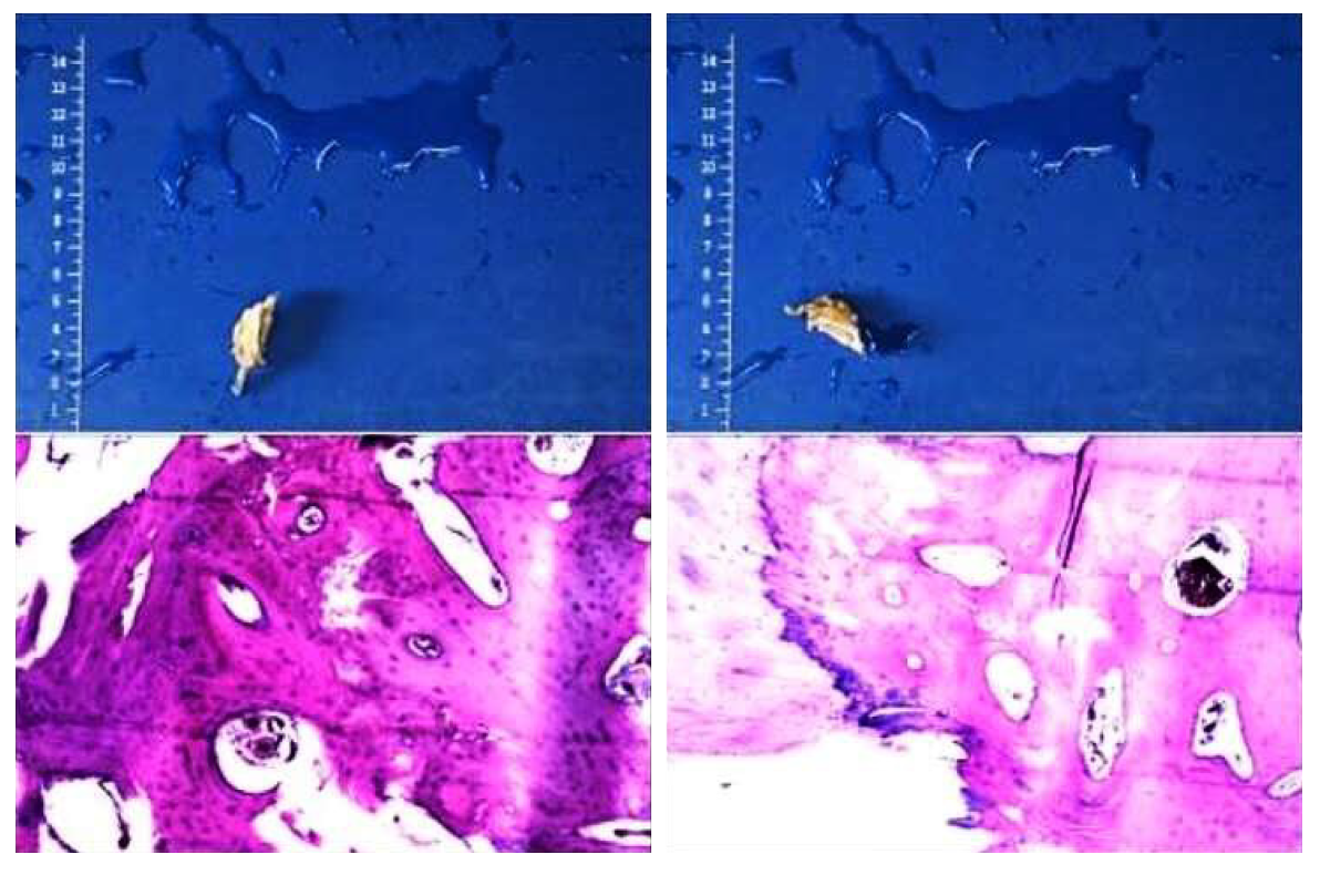
Figure 6 Specimen photos and pathological results.
The Gray-yellow bone tissue seen by the naked eye, is about 2.5 cm × 2.2 cm × 1.3 cm in size, and the mature bone and cartilage tissue seen under microscope staining, is consistent with benign lesions.
- Citation: Cai MD, Zhang HF, Fan YG, Su XJ, Xia L. Obturator nerve impingement caused by an osteophyte in the sacroiliac joint: A case report. World J Clin Cases 2021; 9(5): 1168-1174
- URL: https://www.wjgnet.com/2307-8960/full/v9/i5/1168.htm
- DOI: https://dx.doi.org/10.12998/wjcc.v9.i5.1168


