Copyright
©The Author(s) 2021.
World J Clin Cases. Feb 16, 2021; 9(5): 1156-1167
Published online Feb 16, 2021. doi: 10.12998/wjcc.v9.i5.1156
Published online Feb 16, 2021. doi: 10.12998/wjcc.v9.i5.1156
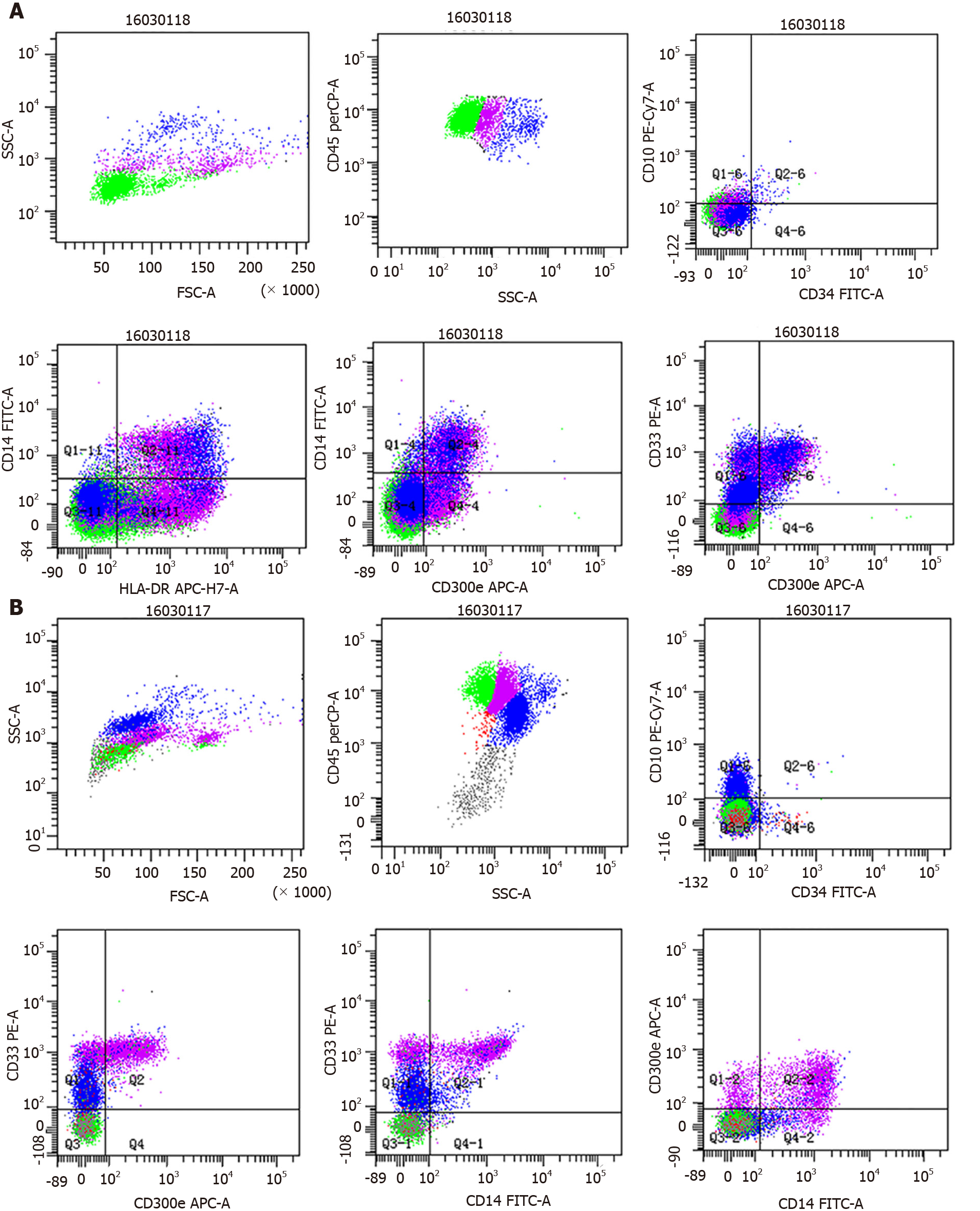
Figure 1 Flow cytometry analyses of patient bronchoalveolar lavage and bone marrow.
A: Bronchoalveolar lavage fluid samples were analyzed via flow cytometry, revealed an increased proportion of monocytes (12.4%, purple), CD33+ CD14+ CD300e+ mature monocytes accounted for approximately 4% of nucleated cells, and CD33+ CD14- CD300e+ partially immature monocyte accounted for 8.4% of nucleated cells; B: Flow cytometry studies of bone marrow samples revealed an increased proportion of monocytes (purple) that accounted for 44% of nucleated cells, and CD33bright CD14- CD300e+ immature monocytes accounted for approximately 7.4% of nucleated cells.
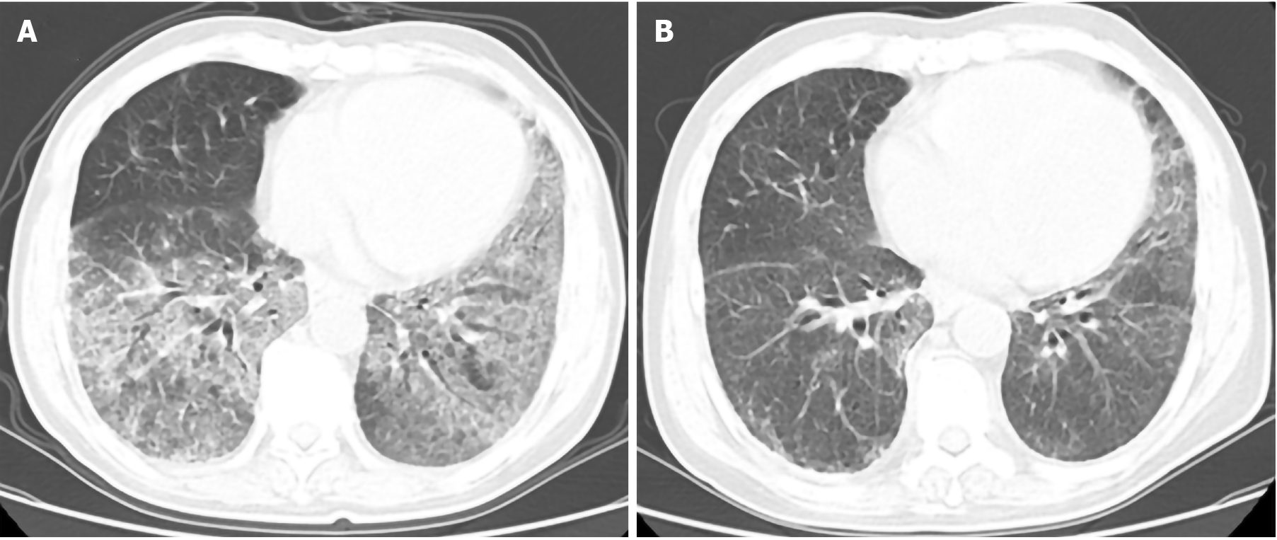
Figure 2 Lung computed tomography findings before and after treatment.
A: Computed tomography scans revealed bilateral regions of ground-glass opacity in the lungs before treatment; B: Computed tomography scans revealed good responses to treatment.
- Citation: Chen C, Huang XL, Gao DQ, Li YW, Qian SX. Chronic myelomonocytic leukemia-associated pulmonary alveolar proteinosis: A case report and review of literature. World J Clin Cases 2021; 9(5): 1156-1167
- URL: https://www.wjgnet.com/2307-8960/full/v9/i5/1156.htm
- DOI: https://dx.doi.org/10.12998/wjcc.v9.i5.1156


