Copyright
©The Author(s) 2021.
World J Clin Cases. Feb 16, 2021; 9(5): 1096-1102
Published online Feb 16, 2021. doi: 10.12998/wjcc.v9.i5.1096
Published online Feb 16, 2021. doi: 10.12998/wjcc.v9.i5.1096
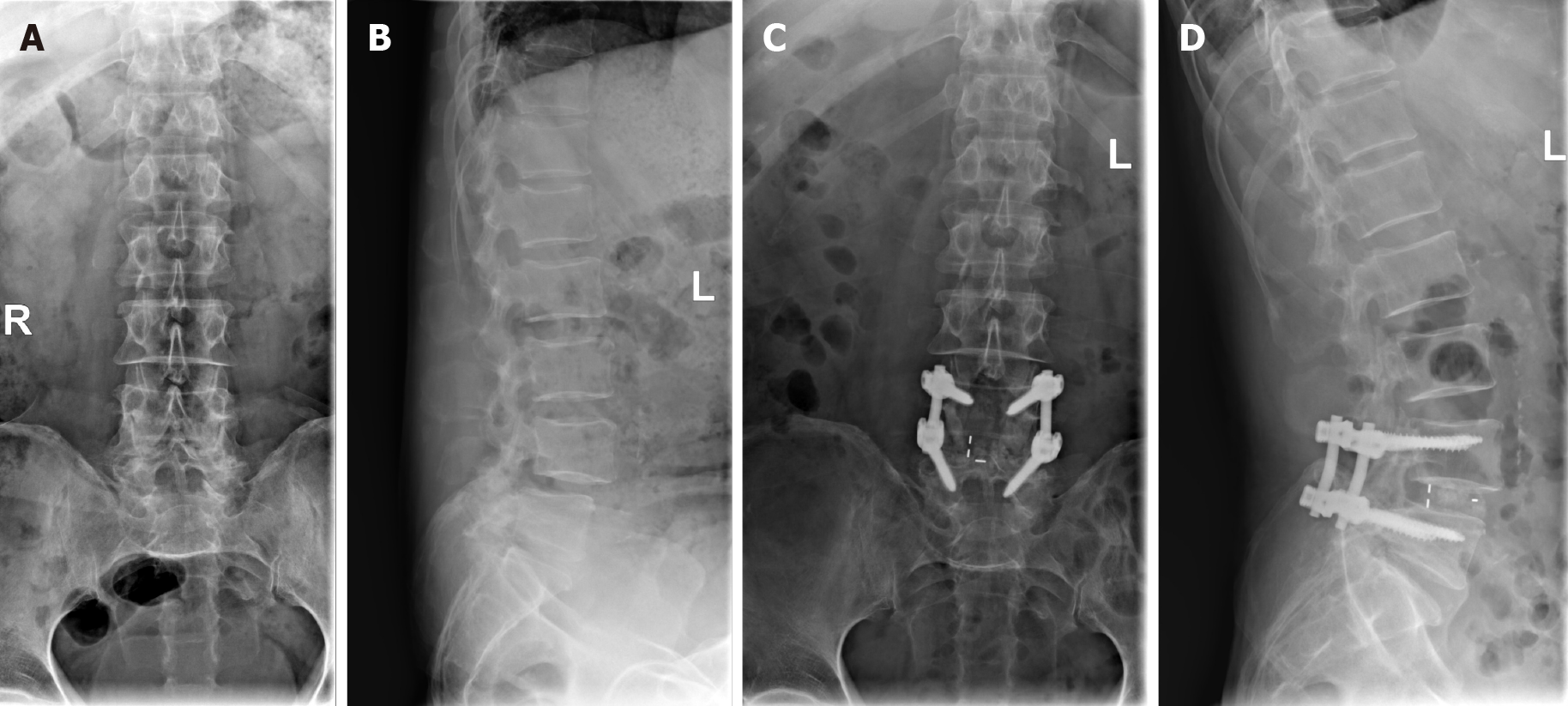
Figure 1 X-rays showed degenerative changes of the lumbar spine and the L4 vertebral body had slipped forward slightly.
Preoperative anterior-posterior (A) and lateral (B) radiographs of the spine demonstrating degenerative changes of the lumbar spine and that the L4 vertebral body had slipped forward slightly. Postoperative anterior-posterior (C) and lateral (D) radiographs of the spine demonstrating the L4-5 instrumented fusion with bilateral segmental pedicle screws-rods fixation and interbody cage devices.
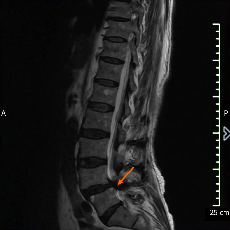
Figure 2 Sagittal view of T2 weighted magnetic resonance imaging.
The arrow shows severe canal stenosis at L4/5.
- Citation: Xu DF, Wu B, Wang JX, Yu J, Xie JX. Severe lumbar spinal stenosis combined with Guillain-Barré syndrome: A case report. World J Clin Cases 2021; 9(5): 1096-1102
- URL: https://www.wjgnet.com/2307-8960/full/v9/i5/1096.htm
- DOI: https://dx.doi.org/10.12998/wjcc.v9.i5.1096


