Copyright
©The Author(s) 2021.
World J Clin Cases. Feb 16, 2021; 9(5): 1079-1086
Published online Feb 16, 2021. doi: 10.12998/wjcc.v9.i5.1079
Published online Feb 16, 2021. doi: 10.12998/wjcc.v9.i5.1079
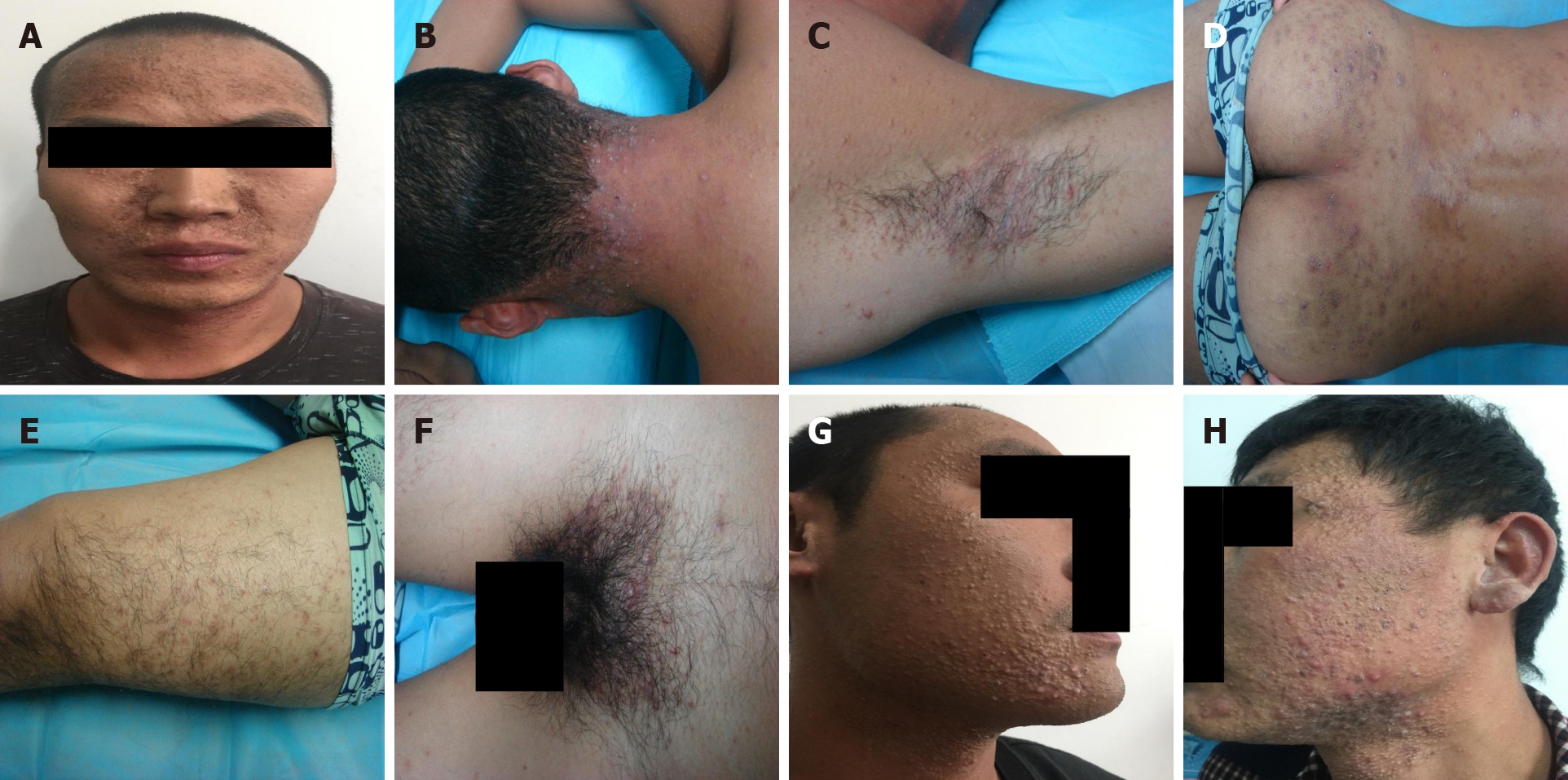
Figure 1 Clinical presentations of the three patients.
A-F: Patient 1 showed grey-black changes on the face, and intensive distribution of yellow to brownish, miliary to mung bean-sized papules and nodules over the neck, armpit, lower limb, hips, and perineum. Some lesions were covered with pus plugs and blackheads, while some formed rashes with confluent patches; G: Patient 2 showed intensive distribution of papules and nodules of varying sizes on the face and neck; and some lesions were covered with pus plugs; H: Patient 3 showed intensive distribution of papules and nodules of varying sizes on the face and neck; and some lesions were covered with pus plugs.
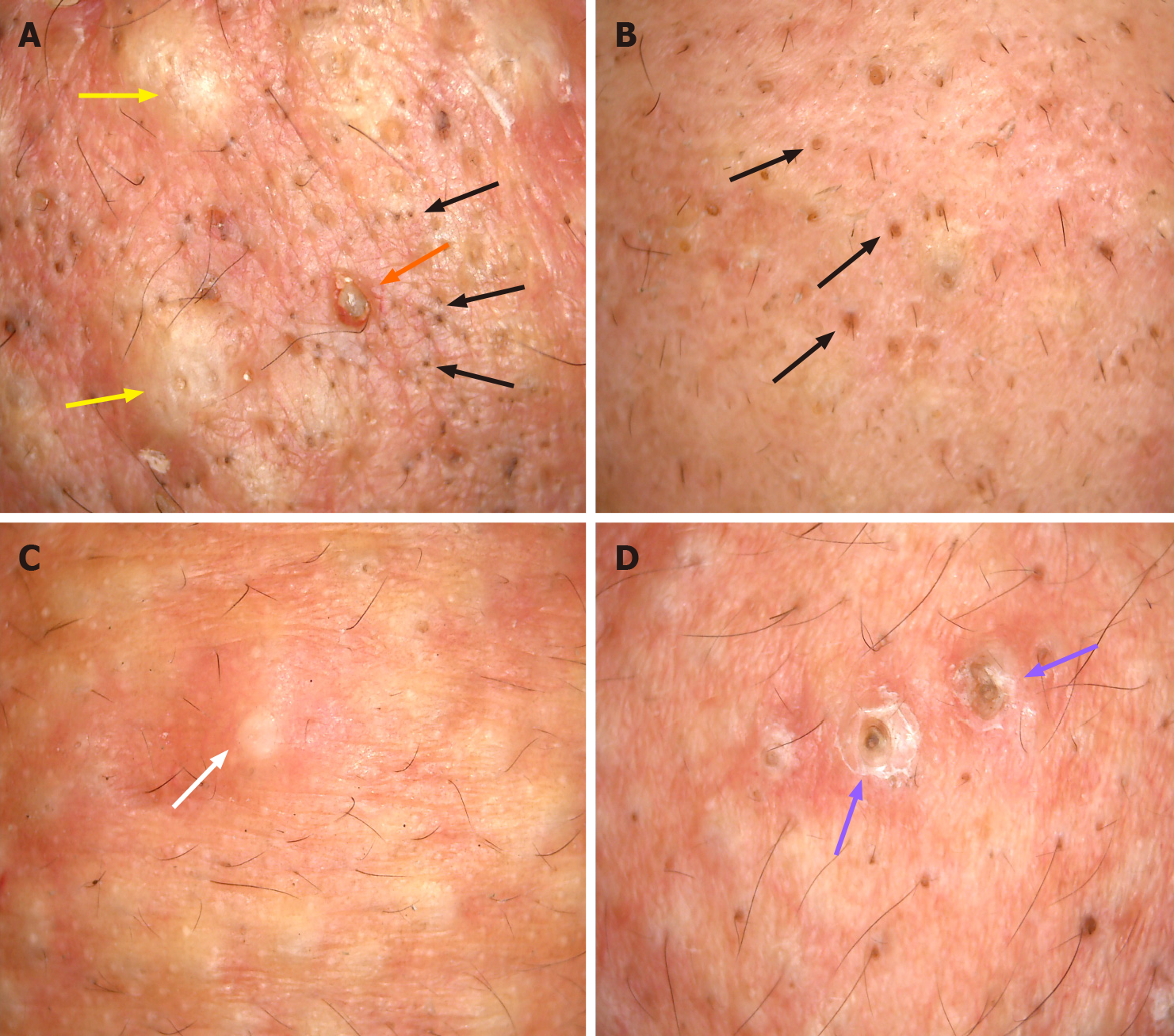
Figure 2 Dermoscopic manifestations of the head and face skin of the three patients (× 30).
A-D: Light red background, with numerous blackheads (black arrows) (A and B), slightly larger solid black and brown plugs (purple arrows) (D), yellow-white inflammatory papules (yellow arrows) (A), pus (white arrow) (C), inflammatory erythema, and focally branched atypical blood vessels (orange arrow) (A).
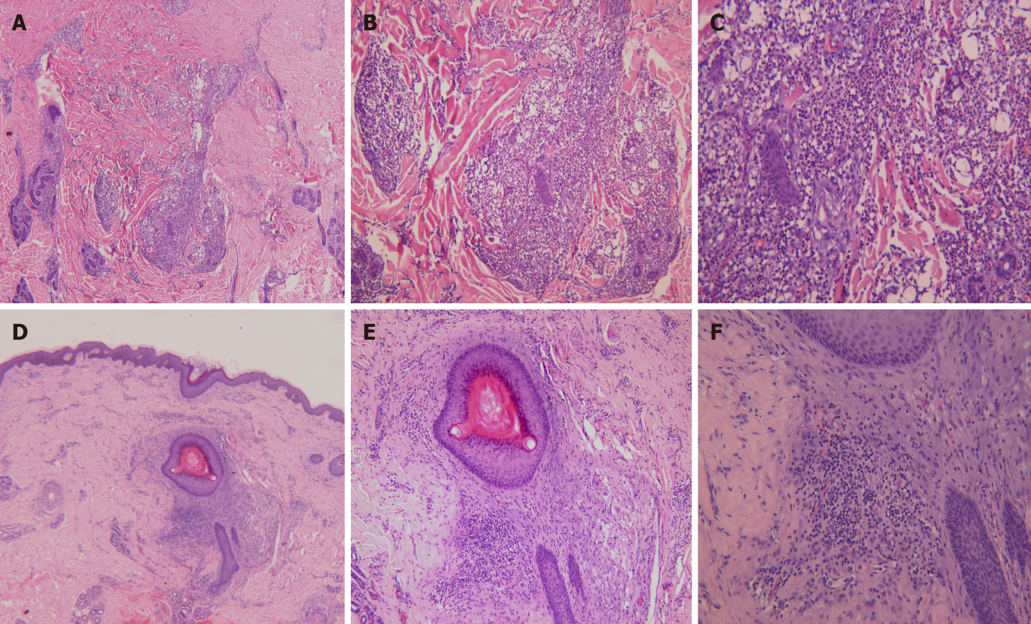
Figure 3 Histopathological staining of the skin eruption.
A-C: Histopathological images of the back skin eruption revealing hyperkeratinization, follicular keratotic plugs, focal epidermal hyperproliferation, vasodilatation and hyperemia in the dermis, destruction of hair follicles and sebaceous glands, and massive perifollicular neutrophil-dominant inflammatory infiltration, suggesting folliculitis and perifolliculitis (A, hematoxylin-eosin [HE] × 40; B, HE × 100; C, HE × 200); D-F: Histopathological images of the ear skin eruption revealing fairly normal epidermis and perifollicular lymphocyte-dominant inflammatory infiltration, suggesting perifolliculitis (D, HE × 40; E, HE × 100; F, HE × 200).
- Citation: Ma Y, Cao X, Zhang L, Zhang JY, Qiao ZS, Feng WL. Neuropathy and chloracne induced by 3,5,6-trichloropyridin-2-ol sodium: Report of three cases. World J Clin Cases 2021; 9(5): 1079-1086
- URL: https://www.wjgnet.com/2307-8960/full/v9/i5/1079.htm
- DOI: https://dx.doi.org/10.12998/wjcc.v9.i5.1079


