Copyright
©The Author(s) 2021.
World J Clin Cases. Feb 6, 2021; 9(4): 983-991
Published online Feb 6, 2021. doi: 10.12998/wjcc.v9.i4.983
Published online Feb 6, 2021. doi: 10.12998/wjcc.v9.i4.983
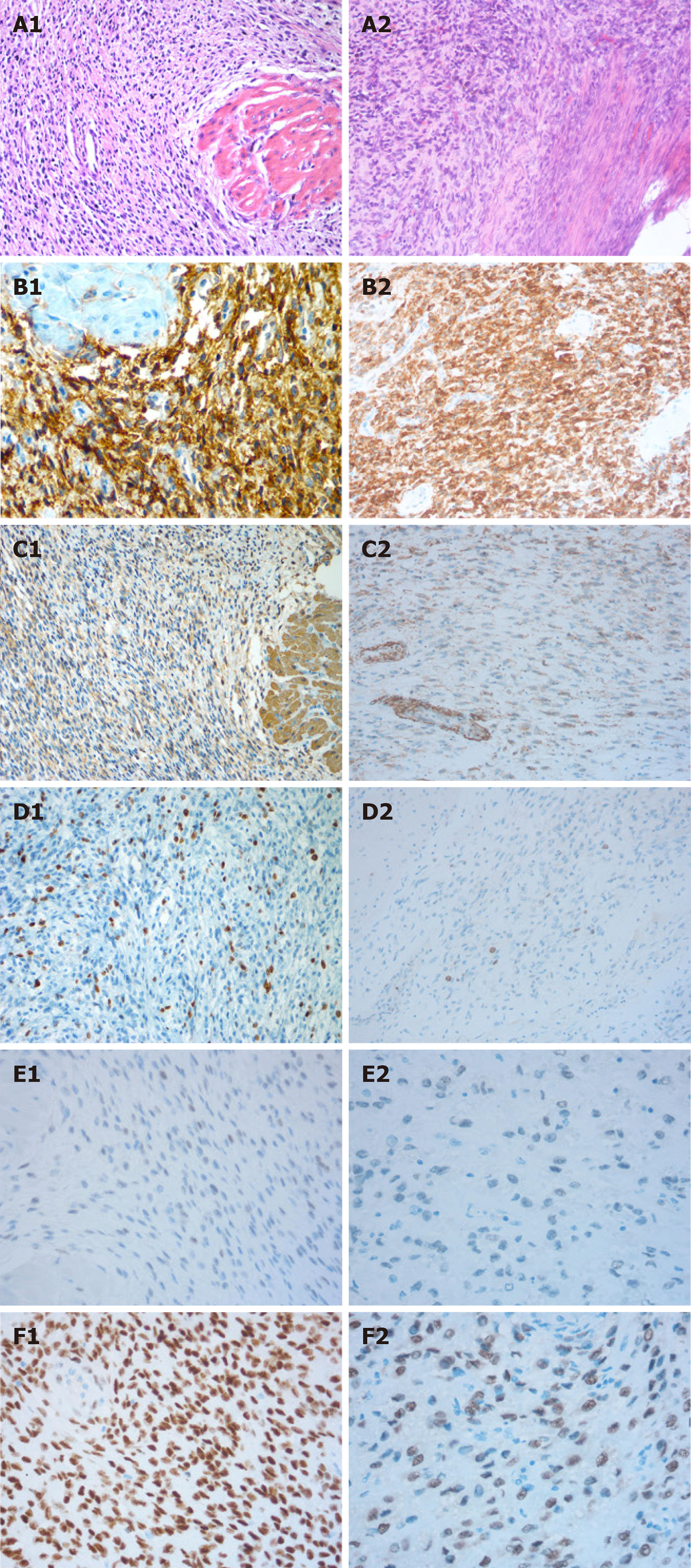
Figure 1 Microscopic and immunohistochemical features of the first (A1-F1) and second (A2-F2) resected tissues.
A: Extensive permeation of the myometrium as irregular islands (hematoxylin-eosin, HE × 200); B: Strong CD10 positivity (brown) (B1) and moderate CD10 positivity (B2) (× 200); C: Smooth muscle actin (SMA) negativity in tumor tissue but positivity in the myometrium is positive (C1) and SMA negativity (C2) (× 200); D: Ki-67 (+;10%) (D1) and Ki-67 (+; 10-15%) (D2) (× 200); E: Estrogen receptor (ER) positivity in 5% of cells (E1 and E2) (× 400) (E2); F: Progestin receptor (PR) positivity in 90% (F1) and 50% (F2) of cells (× 400).
- Citation: Gu YZ, Duan NY, Cheng HX, Xu LQ, Meng JL. Fertility-sparing surgeries without adjuvant therapy through term pregnancies in a patient with low-grade endometrial stromal sarcoma: A case report. World J Clin Cases 2021; 9(4): 983-991
- URL: https://www.wjgnet.com/2307-8960/full/v9/i4/983.htm
- DOI: https://dx.doi.org/10.12998/wjcc.v9.i4.983


