Copyright
©The Author(s) 2021.
World J Clin Cases. Feb 6, 2021; 9(4): 976-982
Published online Feb 6, 2021. doi: 10.12998/wjcc.v9.i4.976
Published online Feb 6, 2021. doi: 10.12998/wjcc.v9.i4.976
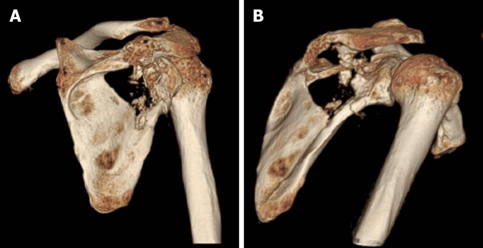
Figure 1 Three-dimensional computed tomography reconstruction.
A and B: Osteolytic destructive lesion in the right scapular bone.
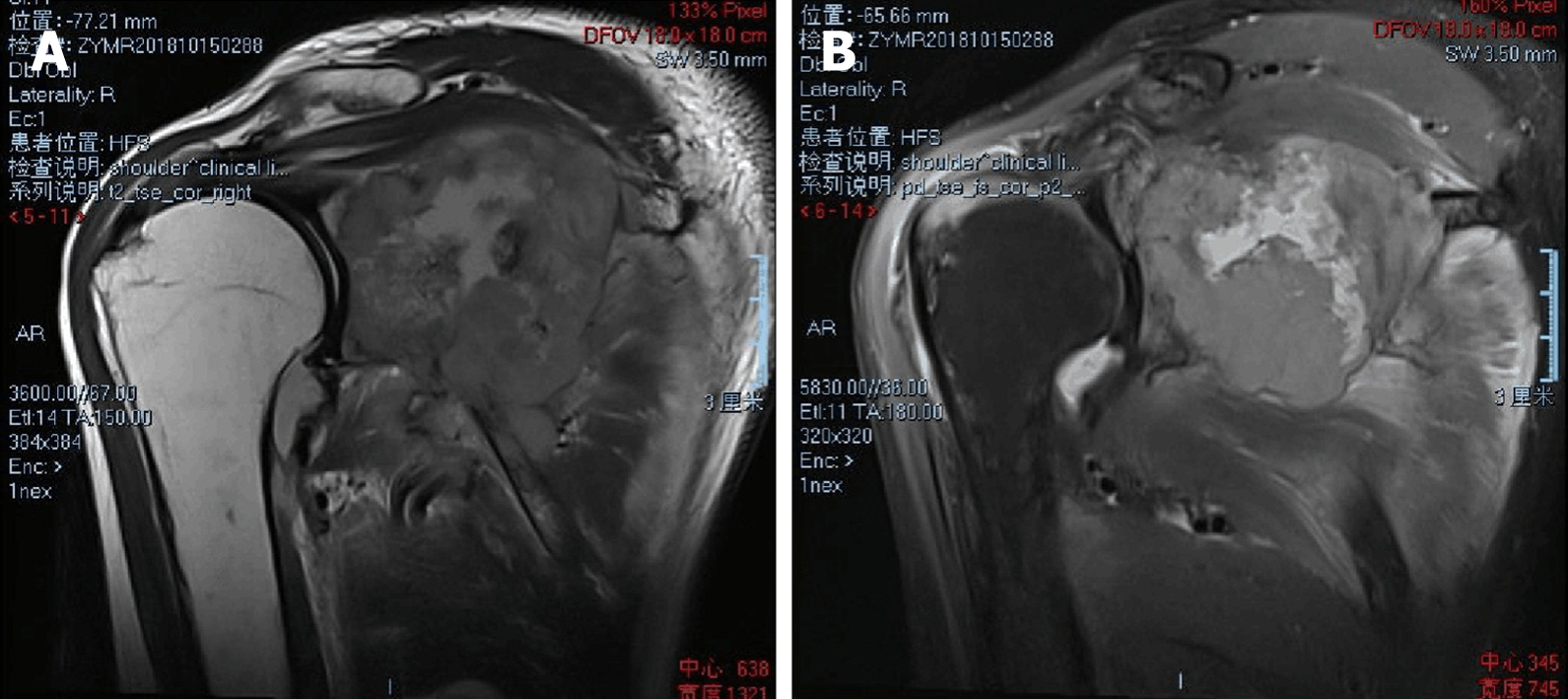
Figure 2 Magnetic resonance imaging A (t2-tse) and B (pd-tse-fs) show an osteolytic destructive lesion in the right scapular bone, invading the surrounding muscles and soft tissues.
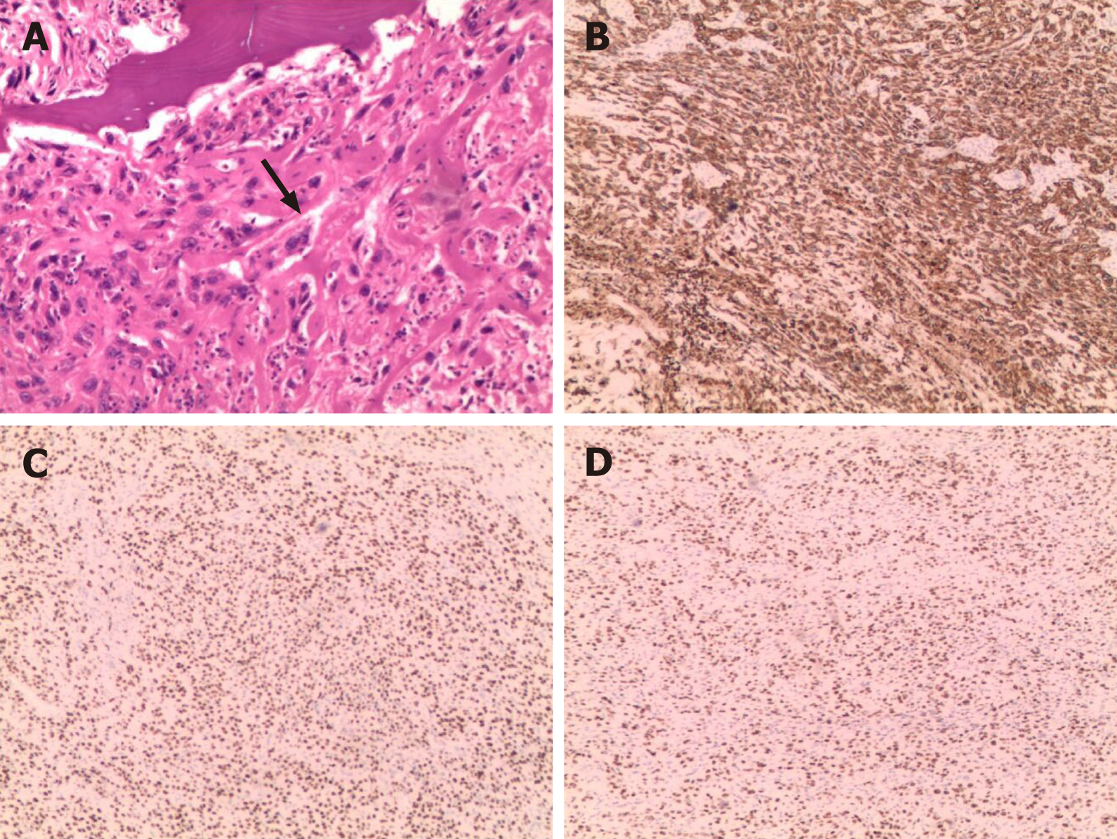
Figure 3 Histological findings.
A: Histopathological examination of the biopsy specimen shows malignant tumor cells in the trabecular bone space but no typical keratin pearls (hematoxylin and eosin stain; original magnification, 100 ×); B-D: Immunohistochemical labeling reveals that the tumor cells are reactive to cytokeratin 5/6, p63, and p40 (original magnification, 40 ×).
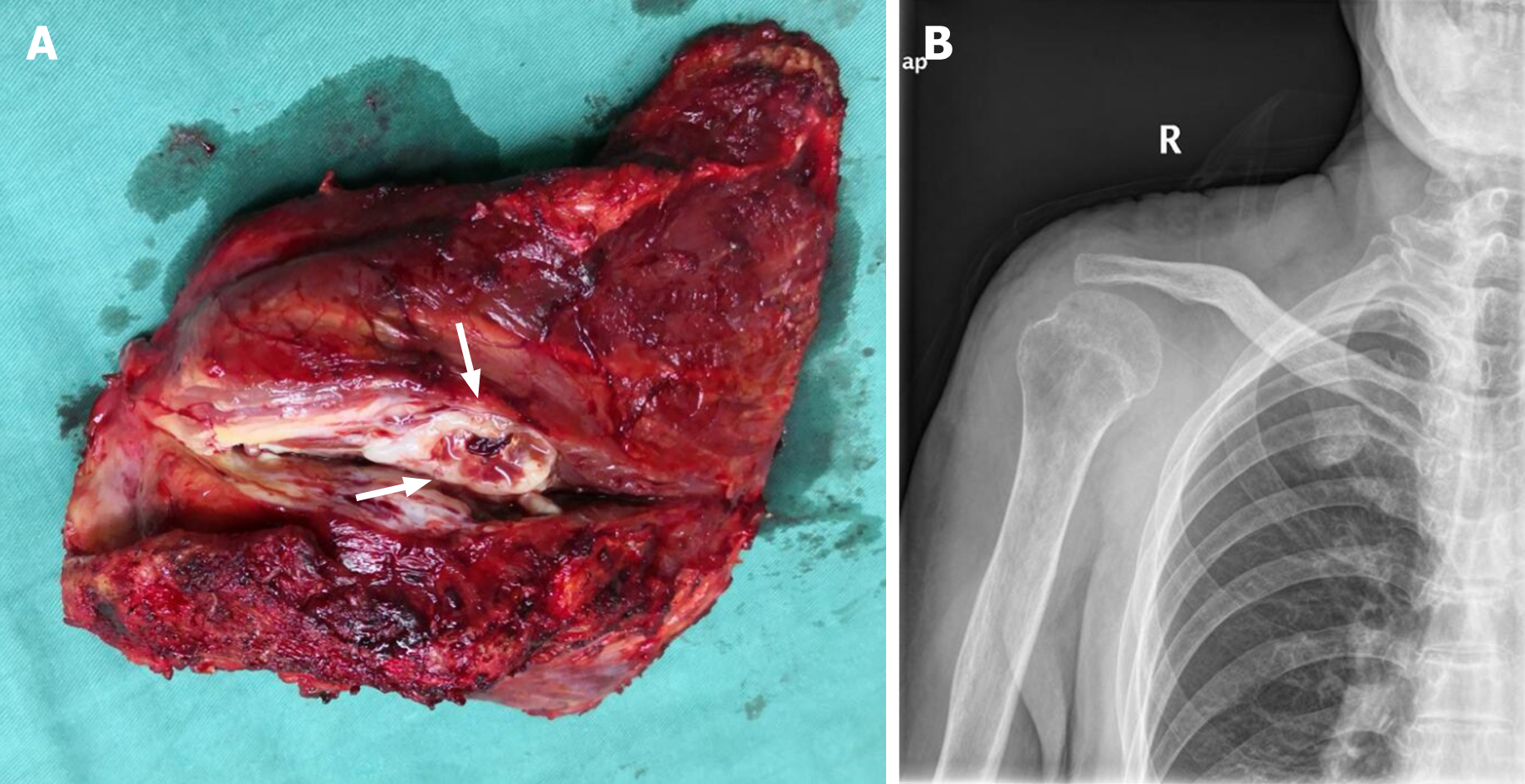
Figure 4 Primary nonkeratinizing squamous cell carcinoma of the right scapular bone.
A: Surgically resected specimen shows a dark red ovoid mass originating from the right scapula that appears creamy-white and soft on cross section; B: The right humeral head, right collarbone, and surrounding muscle were used to reconstruct the resulting defect.
- Citation: Li Y, Zuo JL, Tang JS, Shen XY, Xu SH, Xiao JL. Primary nonkeratinizing squamous cell carcinoma of the scapular bone: A case report. World J Clin Cases 2021; 9(4): 976-982
- URL: https://www.wjgnet.com/2307-8960/full/v9/i4/976.htm
- DOI: https://dx.doi.org/10.12998/wjcc.v9.i4.976


