Copyright
©The Author(s) 2021.
World J Clin Cases. Feb 6, 2021; 9(4): 970-975
Published online Feb 6, 2021. doi: 10.12998/wjcc.v9.i4.970
Published online Feb 6, 2021. doi: 10.12998/wjcc.v9.i4.970
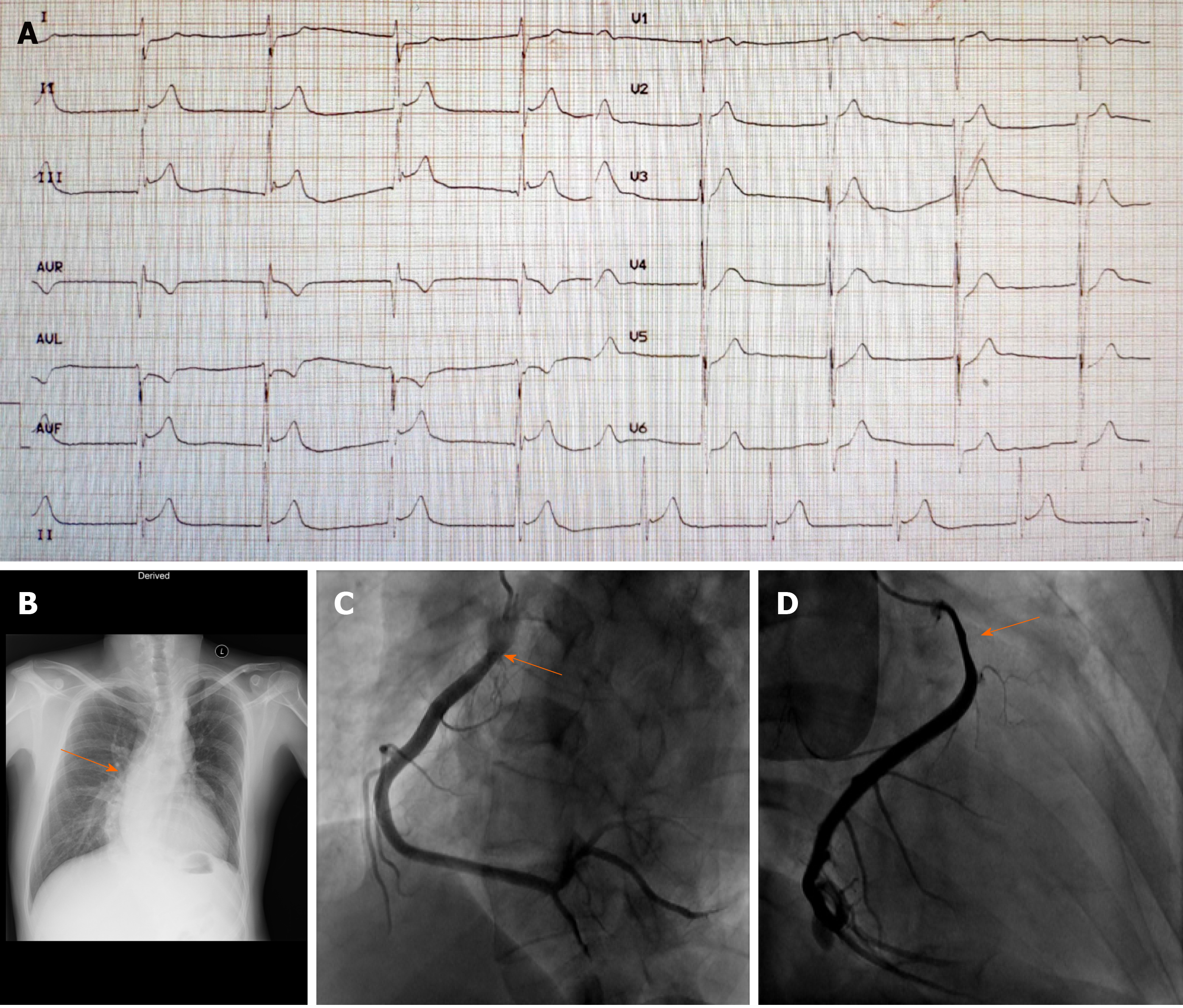
Figure 1 Imaging examinations.
A: Twelve lead electrocardiogram showing ST-segment elevation in leads II, III, and aVF and deep reciprocal ST depression in leads I and aVL; B: Chest X-rays showing scoliosis (arrow); C and D: Coronary angiography showing an eccentric stenosis in the proximal segment of right coronary artery (arrow).
Figure 2 Computed tomography angiography results.
A: Focal aortic intramural thrombosis on axial aortic computed tomography angiography (CTA) (arrow); B: Focal aortic intramural thrombosis on sagittal aortic CTA (arrow); C: Full recovery of the aorta.
- Citation: Zhang YX, Yang H, Wang GS. Acute inferior wall myocardial infarction induced by aortic dissection in a young adult with Marfan syndrome: A case report. World J Clin Cases 2021; 9(4): 970-975
- URL: https://www.wjgnet.com/2307-8960/full/v9/i4/970.htm
- DOI: https://dx.doi.org/10.12998/wjcc.v9.i4.970


