Copyright
©The Author(s) 2021.
World J Clin Cases. Feb 6, 2021; 9(4): 960-969
Published online Feb 6, 2021. doi: 10.12998/wjcc.v9.i4.960
Published online Feb 6, 2021. doi: 10.12998/wjcc.v9.i4.960
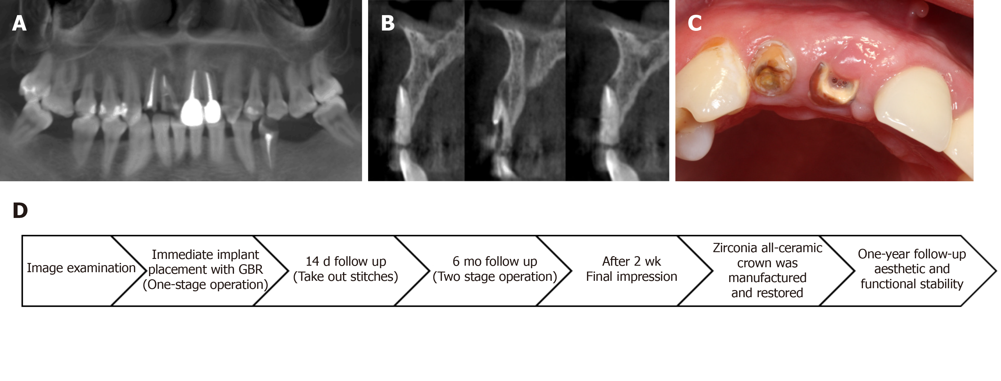
Figure 1 Clinical and radiograph examination before surgery.
A: Intraoral photograph before operation; B: Coronal plane of cone beam computed tomography before operation; C: Sagittal plane of cone beam computed tomography before operation; D: Flow chart timeline of the treatment plan.
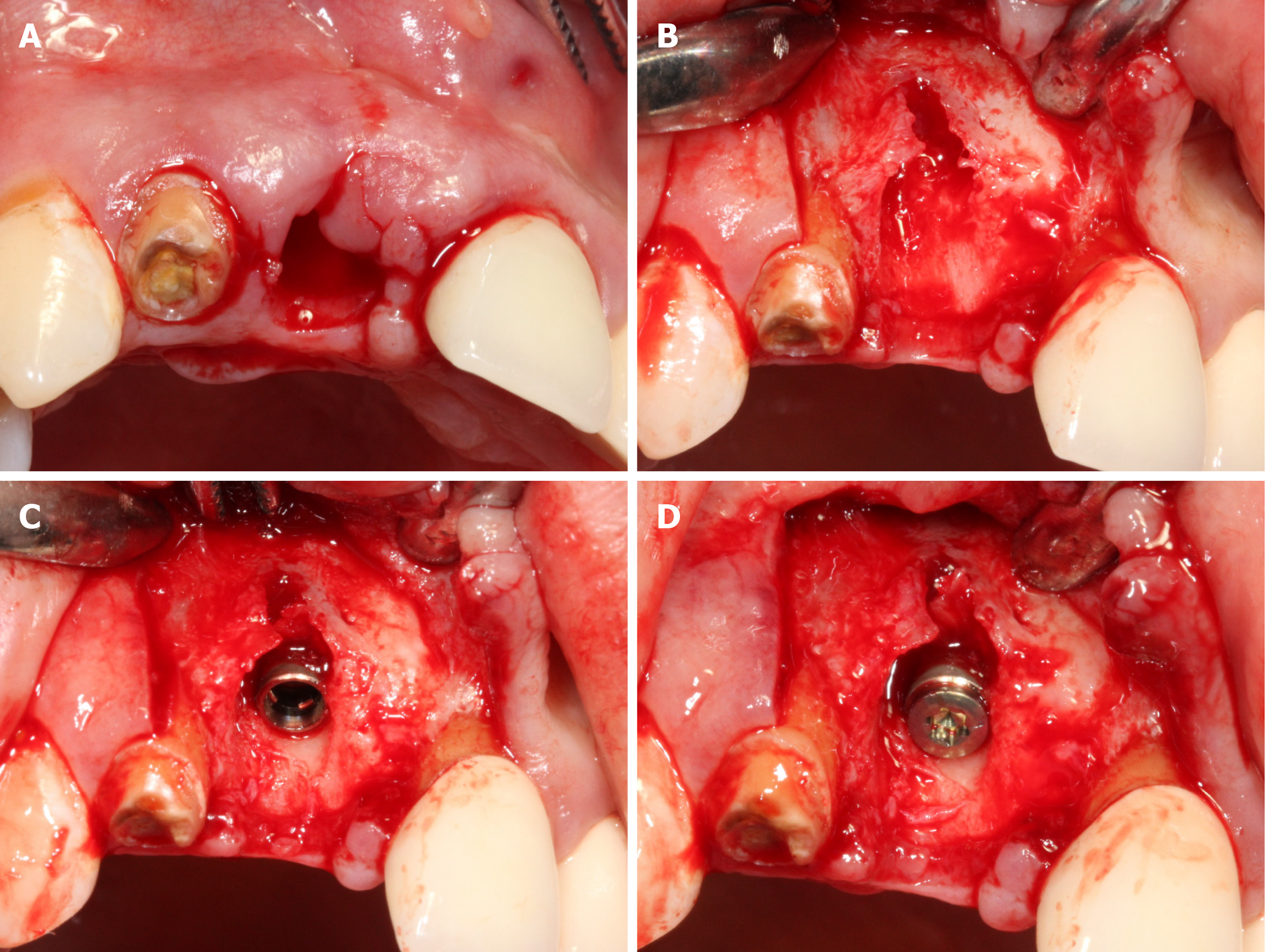
Figure 2 Surgical procedures of immediate implantation.
A: Remove residual root and granulation tissue, then rinse the extraction socket with normal saline; B: Imperfect thin buccal plate with dehiscence can be observed; C: The implant was immediately implanted; D: Cover screw was placed.
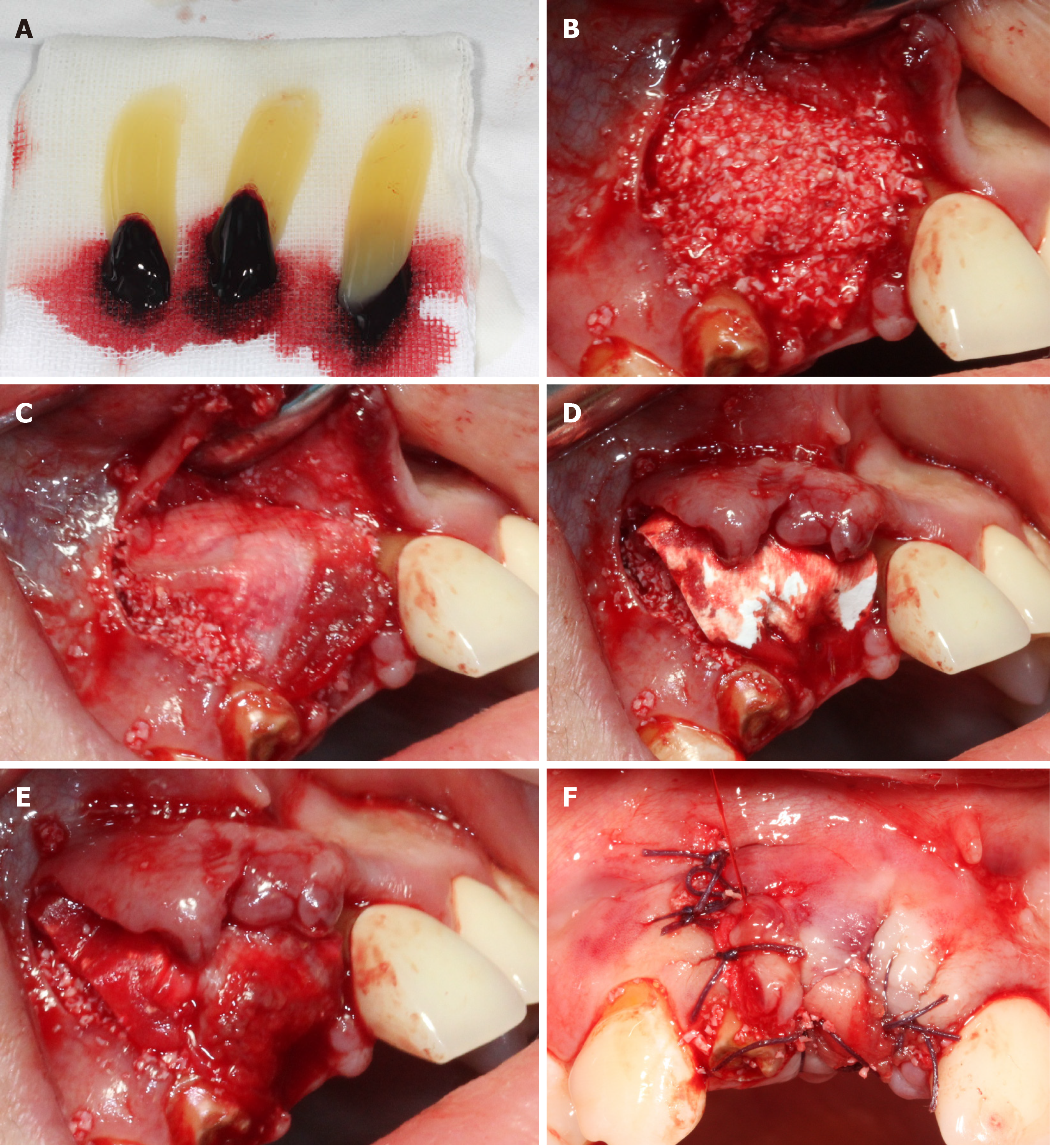
Figure 3 Representational images and illustrations describing each step of covering the barrier membrane.
A: Platelet-rich fibrin (PRF) clots were obtained into the test tube through whole blood centrifuged immediately at 3000 rpm for 10 min; B: PRF clot was mixed with 0.25 g bone substitute (Bio-Oss) then placed on the implant and the bone defect; C: A piece of PRF clot was pressed into a membrane by sterile gauze and placed on the surface of Bio-Oss-PRF mixture; D: Placement of collagen membrane; E: The other PRF membrane was placed on the surface of the collagen membrane; F: Tension-free suture.
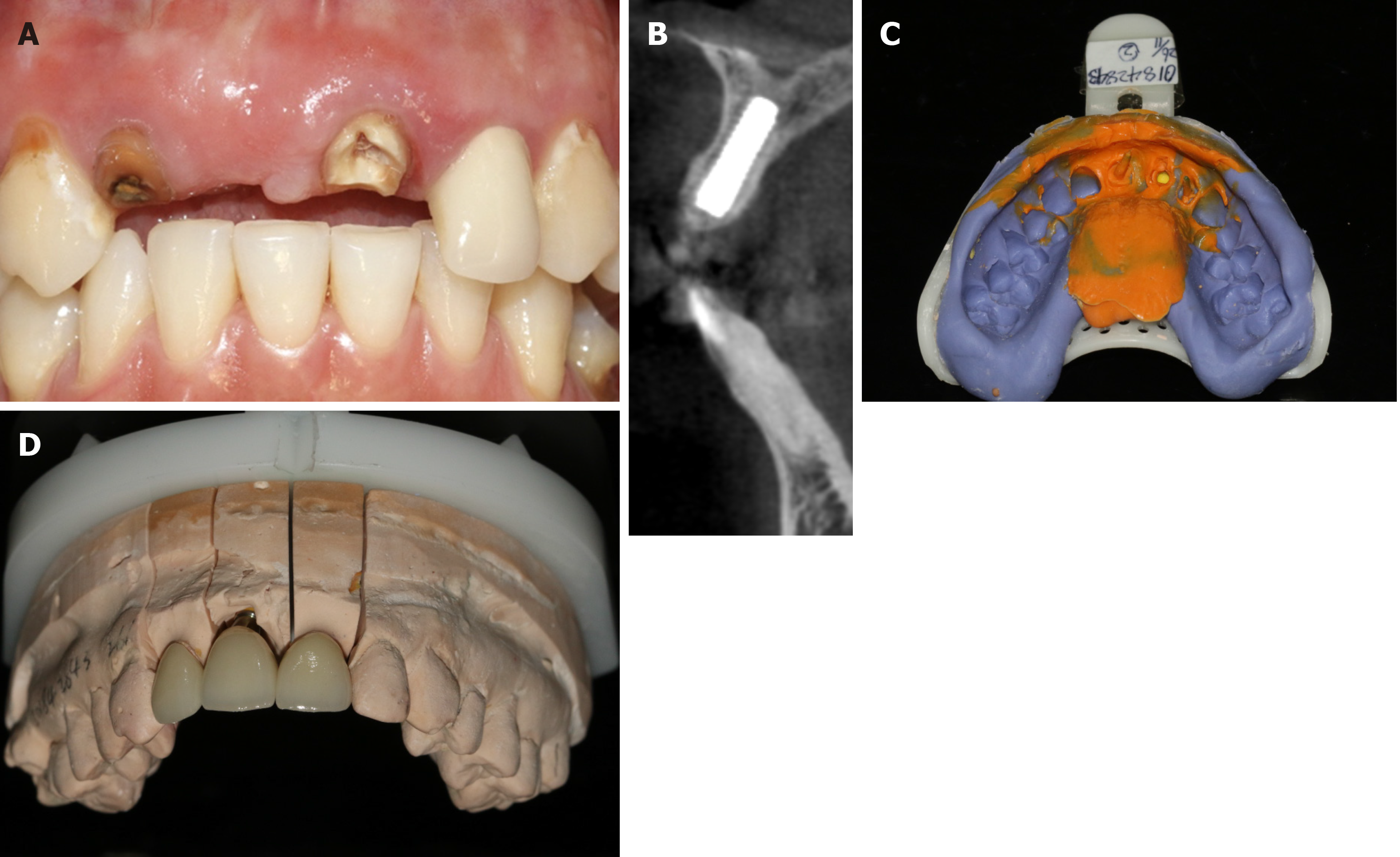
Figure 4 The impression was finished at 24 wk post-surgery.
A: Intraoral photograph; B: Cone beam computed tomography shows the results of bone increment; C: Final impression; D: Crowns were simulated in a gypsum model.
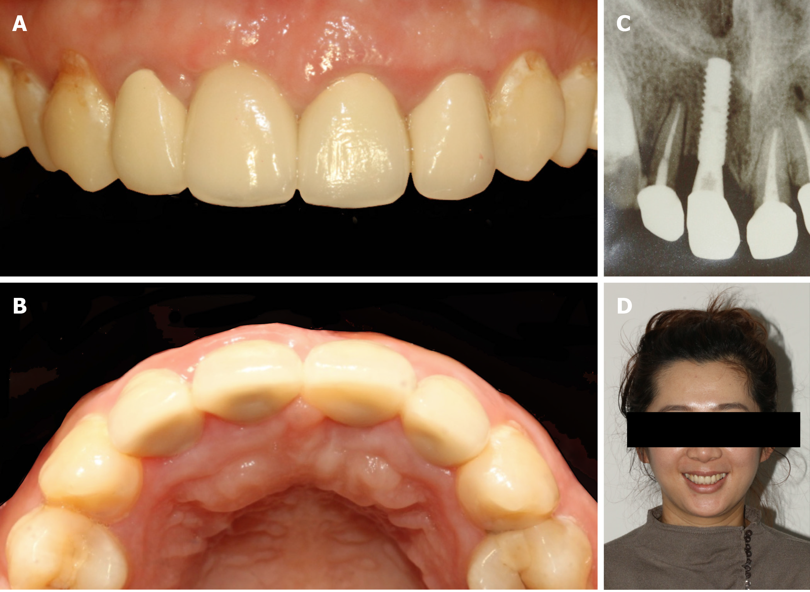
Figure 5 Final restoration crown and X-ray examination.
A: Cementation of a full-ceramic crown; B: Full formed skeletal arch contour; C: All-ceramic crowns of the implant and adjacent teeth well positioned; D: Patient smiling with satisfaction.
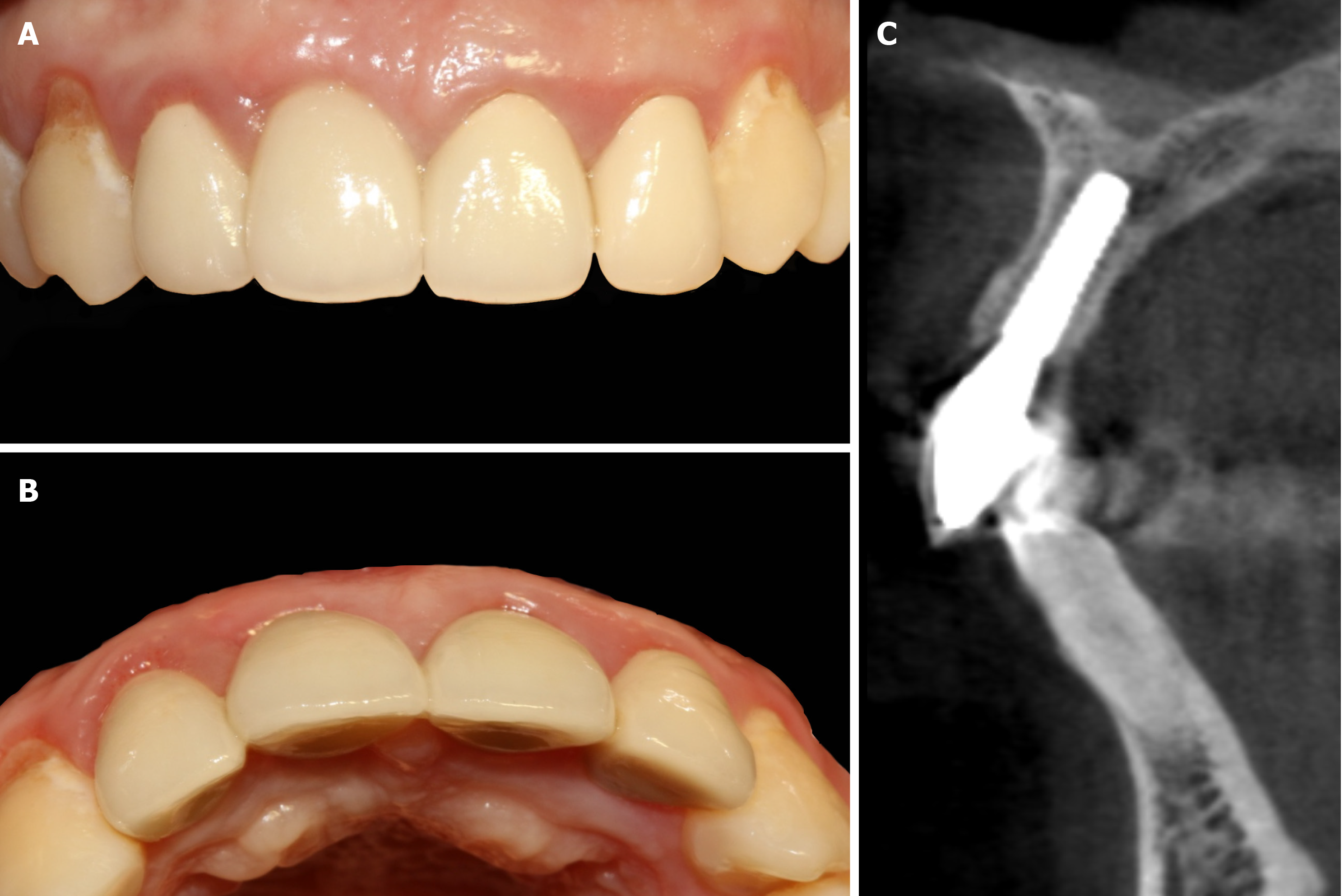
Figure 6 Twelve-month follow-up after restoration.
A: Stable soft tissue around the implant; B: Maintenance of good arch contour; C: No obvious absorption of the labial and palatal bone plates.
- Citation: Fang J, Xin XR, Li W, Wang HC, Lv HX, Zhou YM. Immediate implant placement in combination with platelet rich-fibrin into extraction sites with periapical infection in the esthetic zone: A case report and review of literature. World J Clin Cases 2021; 9(4): 960-969
- URL: https://www.wjgnet.com/2307-8960/full/v9/i4/960.htm
- DOI: https://dx.doi.org/10.12998/wjcc.v9.i4.960


