Copyright
©The Author(s) 2021.
World J Clin Cases. Feb 6, 2021; 9(4): 951-959
Published online Feb 6, 2021. doi: 10.12998/wjcc.v9.i4.951
Published online Feb 6, 2021. doi: 10.12998/wjcc.v9.i4.951
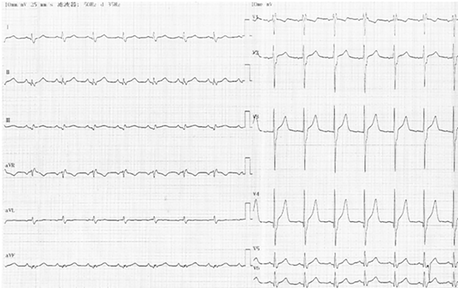
Figure 1 Electrocardiogram at hospital admission.
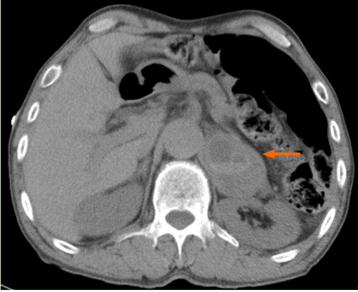
Figure 2 Computed tomography scan revealing an immense mass (marked by arrow) in the left adrenal gland (46.
4 mm × 53.5 mm × 56.0 mm).
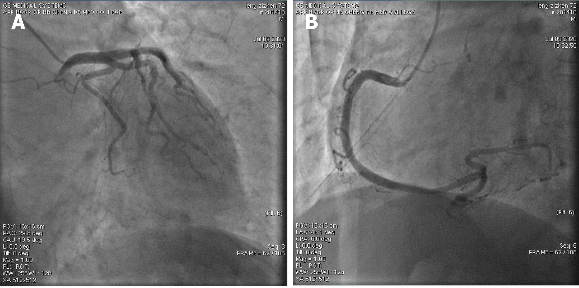
Figure 3 Coronary angiogram showing nonobstructed coronaries.
A: Left coronary arteries; B: Right coronary artery.
- Citation: Shi F, Sun LX, Long S, Zhang Y. Pheochromocytoma as a cause of repeated acute myocardial infarctions, heart failure, and transient erythrocytosis: A case report and review of the literature. World J Clin Cases 2021; 9(4): 951-959
- URL: https://www.wjgnet.com/2307-8960/full/v9/i4/951.htm
- DOI: https://dx.doi.org/10.12998/wjcc.v9.i4.951


