Copyright
©The Author(s) 2021.
World J Clin Cases. Feb 6, 2021; 9(4): 927-934
Published online Feb 6, 2021. doi: 10.12998/wjcc.v9.i4.927
Published online Feb 6, 2021. doi: 10.12998/wjcc.v9.i4.927
Figure 1 Radiographic and pathological outcomes.
A: No abnormalities were observed on X-ray of the shoulder joint; B: Encapsulated effusion was observed in the subacromial bursa upon magnetic resonance imaging; C: Histology of the tissue removed at surgery showed fibrous tissue hyperplasia with localized purulent inflammation changes (white arrow) and neutrophil infiltration (orange arrow). Scale bar: 400 μm.
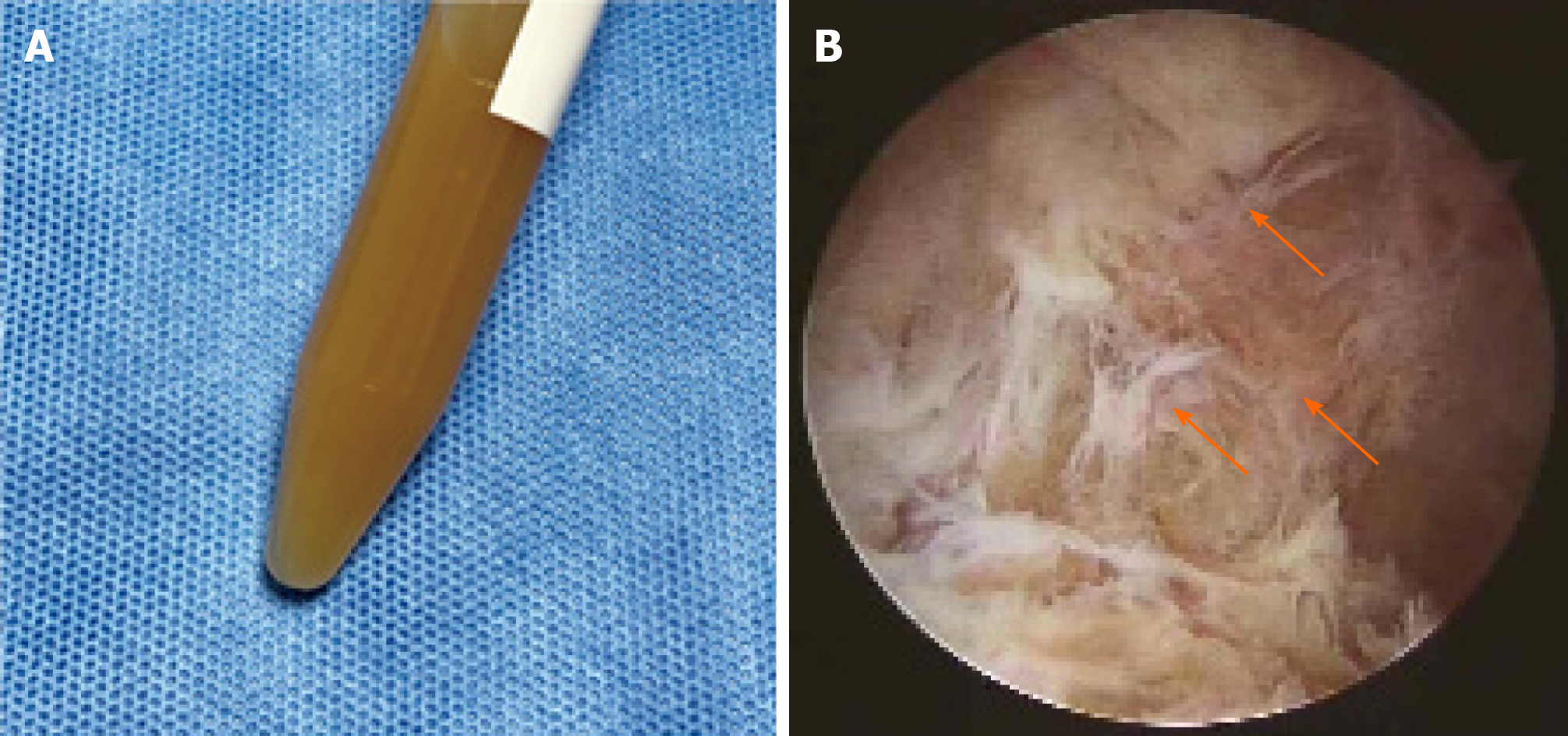
Figure 2 General view of the synovial fluid and subacromial bursa under arthroscopy.
A: The synovial fluid was purulent and turbid; B: The subacromial bursa showed pathological changes of fibrinoid necrosis (arrow) under arthroscopy.
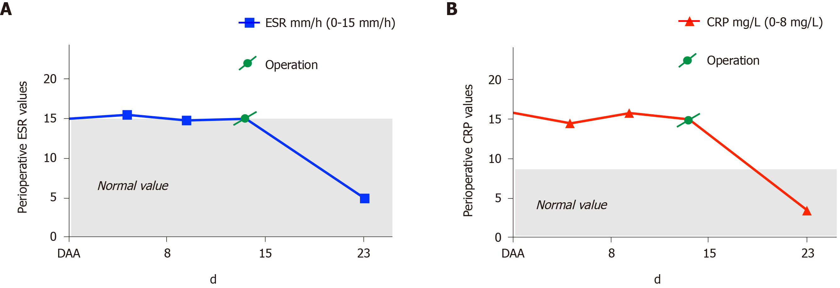
Figure 3 Perioperative erythrocyte sedimentation rate and C-reactive protein values.
A and B: Erythrocyte sedimentation rate (in panel A) and C-reactive protein (in panel B) declined to normal level following the surgical intervention. On both graphs, the x-axis indicates the length of hospital stay. CRP: C-reactive protein; DAA: Days after admission, ESR: Erythrocyte sedimentation rate.
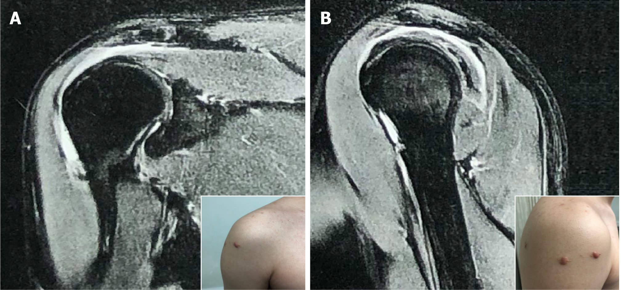
Figure 4 Follow-up imaging evaluation at 4 mo postoperative.
Magnetic resonance imaging showed the effusion of the subacromial bursa to be significantly reduced. A: Frontal view; B: Sagittal view. Corresponding insets show the incisions to have healed well after surgery.
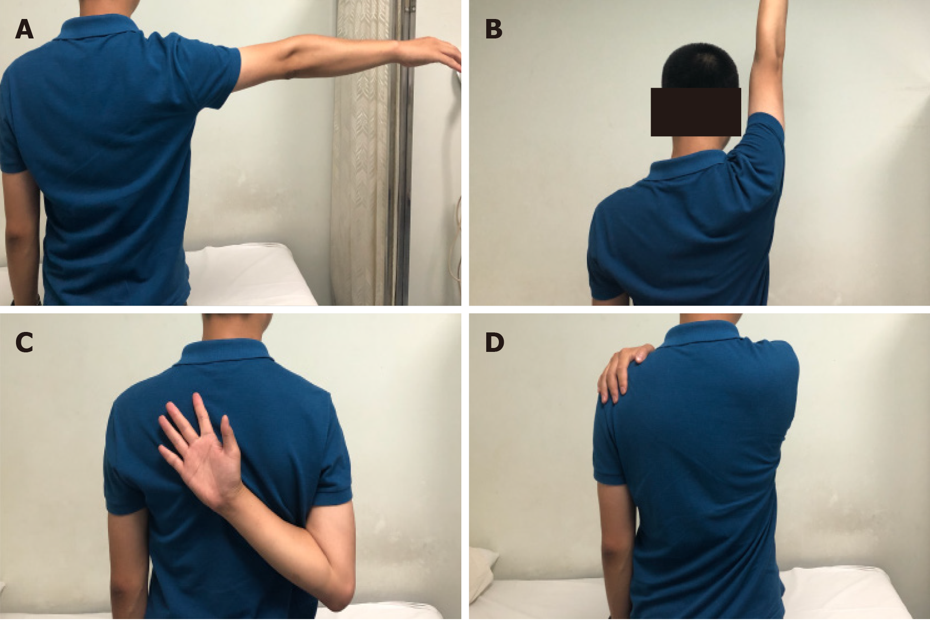
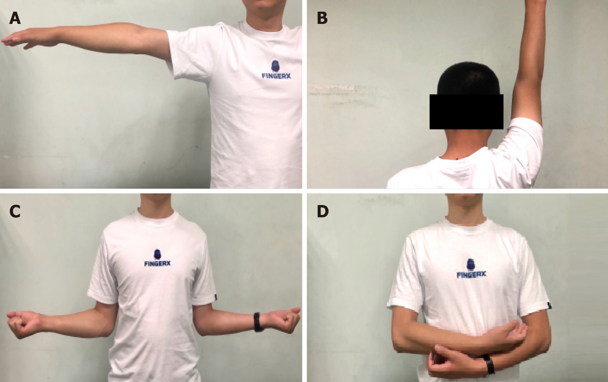
Figure 5 Range of motion of the patient’s right shoulder at 4-mo follow-up.
Function and activity of the shoulder joint recovered well during the first 4 mo after surgery. A: Abduction; B: Flexion C: Internal rotation; D: Adduction.
Figure 6 Range of motion of the patient’s right shoulder at 2-year follow-up.
Function and activity of the shoulder joint were completely normal. A: Abduction; B: Flexion; C: External rotation; D: Internal rotation.
- Citation: Wang FS, Shahzad K, Zhang WG, Li J, Tian K. Atypical presentation of shoulder brucellosis misdiagnosed as subacromial bursitis: A case report . World J Clin Cases 2021; 9(4): 927-934
- URL: https://www.wjgnet.com/2307-8960/full/v9/i4/927.htm
- DOI: https://dx.doi.org/10.12998/wjcc.v9.i4.927


