Copyright
©The Author(s) 2021.
World J Clin Cases. Feb 6, 2021; 9(4): 904-911
Published online Feb 6, 2021. doi: 10.12998/wjcc.v9.i4.904
Published online Feb 6, 2021. doi: 10.12998/wjcc.v9.i4.904
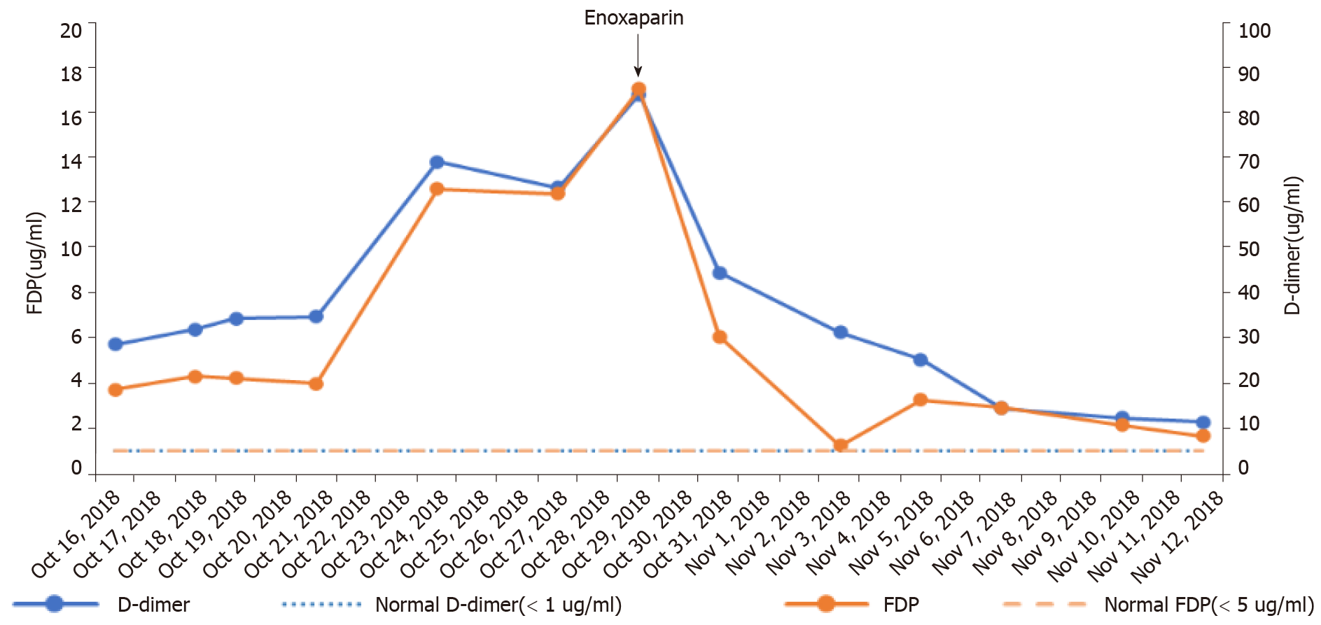
Figure 1 Monitoring of D-dimer and fibrin degradation product levels.
The arrow indicates enoxaparin therapy. FDP: Fibrin degradation product.
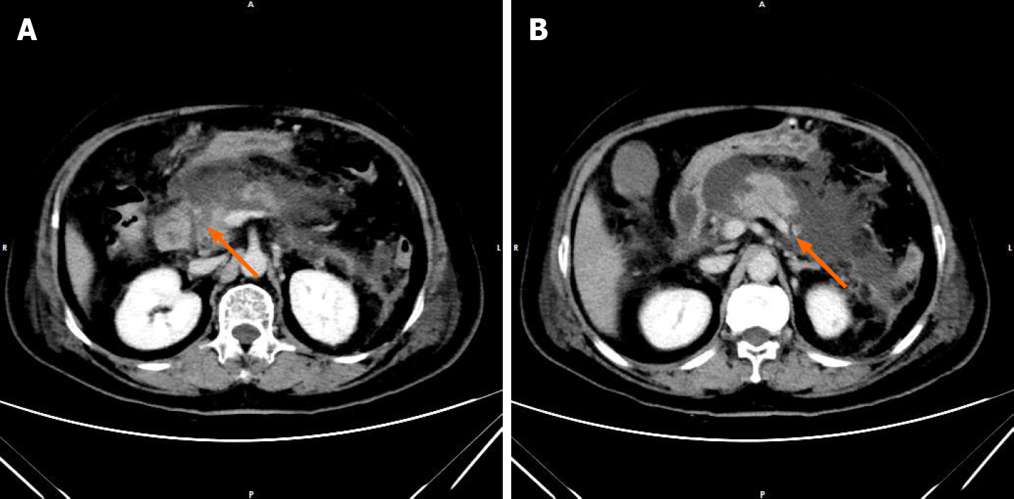
Figure 2 Enhanced computed tomography scan of the abdomen revealed acute necrotizing pancreatitis.
A: Pancreatic head necrosis (arrow); B: Pancreatic body necrosis with accumulation of peripancreatic fluid (arrow).
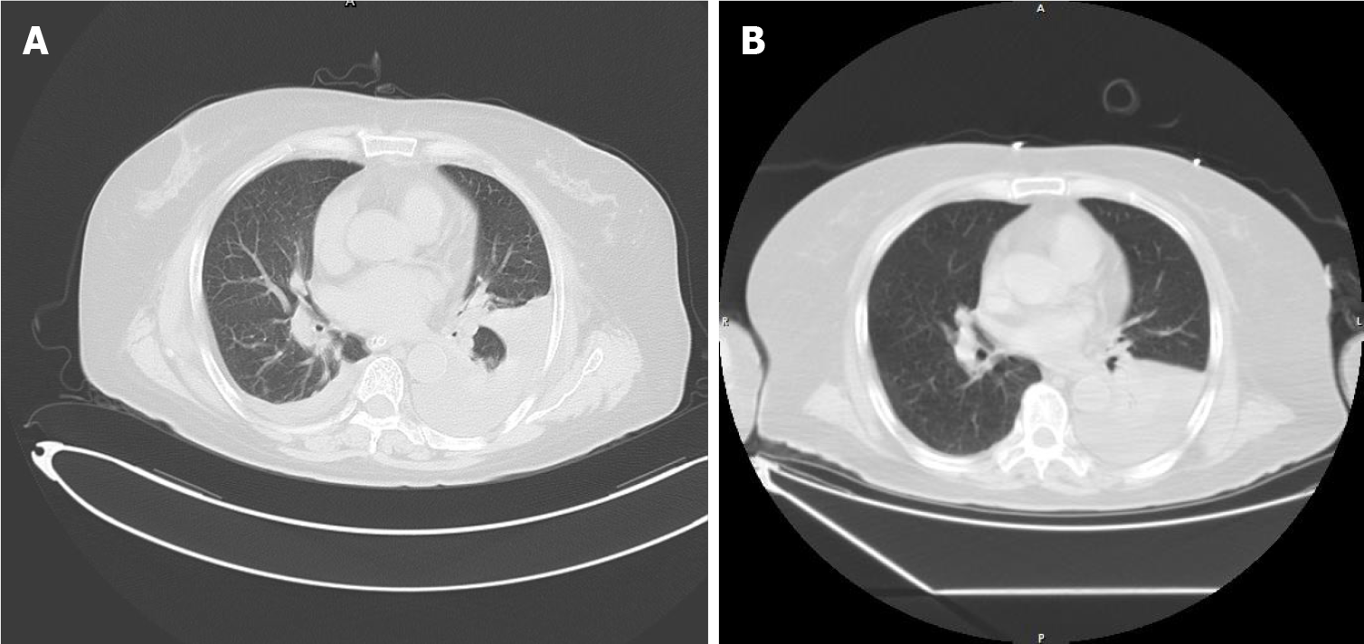
Figure 3 Computed tomography findings.
A computed tomography scan of the chest revealed left pleural effusion, external pressure atelectasis of left lower lobe (lung window) A: Image at admission; B: Image during pulmonary embolism.
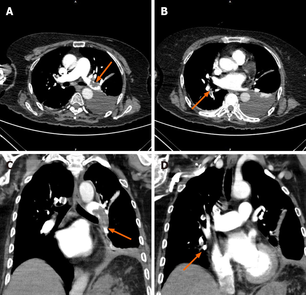
Figure 4 Computed tomography angiography findings.
Computed tomography angiography of the chest revealed pulmonary embolus (the left main pulmonary artery and multiple branches of the left and right pulmonary arteries). A: Embolus of the left main pulmonary artery (arrow); B: Embolus of the right pulmonary artery branch (arrow); C: A large number of emboli in the main trunk and lower lobe of the left pulmonary artery in coronal view of chest-3D slab image (arrow); D: Embolus in branches of the right pulmonary arteries in coronal view of chest-3D slab image (arrow).
- Citation: Fu XL, Liu FK, Li MD, Wu CX. Acute pancreatitis with pulmonary embolism: A case report. World J Clin Cases 2021; 9(4): 904-911
- URL: https://www.wjgnet.com/2307-8960/full/v9/i4/904.htm
- DOI: https://dx.doi.org/10.12998/wjcc.v9.i4.904


