Copyright
©The Author(s) 2021.
World J Clin Cases. Feb 6, 2021; 9(4): 898-903
Published online Feb 6, 2021. doi: 10.12998/wjcc.v9.i4.898
Published online Feb 6, 2021. doi: 10.12998/wjcc.v9.i4.898
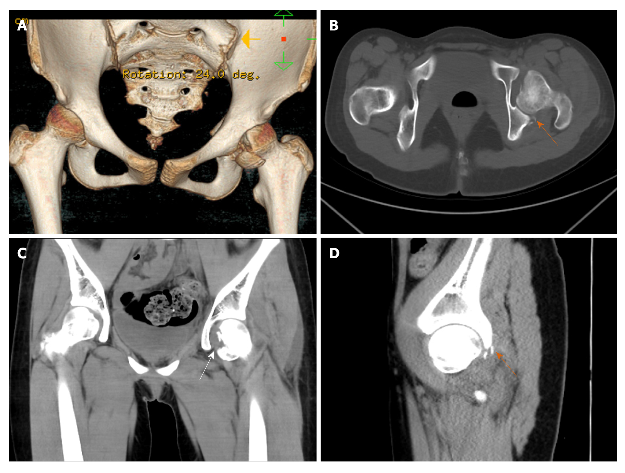
Figure 1 Preoperative computed tomography scans of the hip.
A: Three-dimensional computed tomography scan showed femoral head fracture; B and D: Axial and sagittal computed tomography scans showed the posterior acetabulum with slight displacement (orange arrow); C: Coronal computed tomography scan showed the femoral head fracture extending superior to the fovea centralis (white arrow).
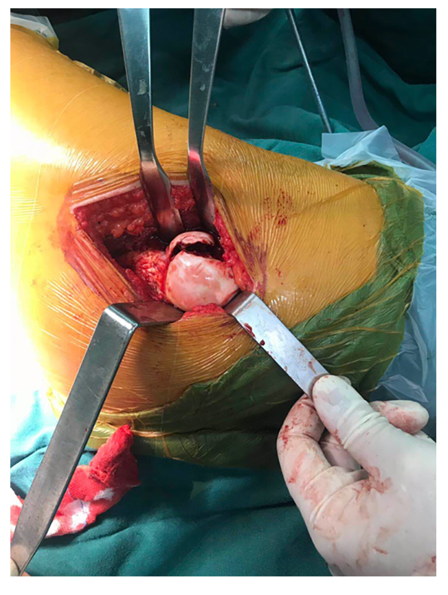
Figure 2 Intraoperative picture showing a major fragment in the anterior and inferior of the femoral head.
The patient was in lateral position.
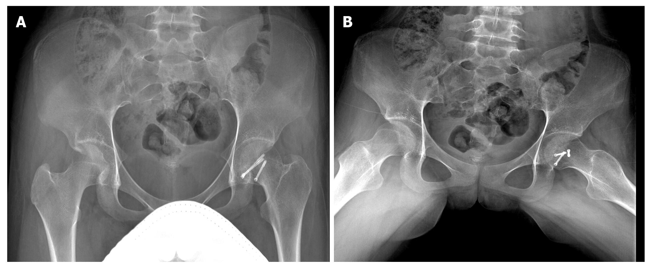
Figure 3 Pelvic radiographs 1 year after operation.
A and B: Anteroposterior x-ray images showed union of the fractures with no femoral head necrosis.
- Citation: Liu Y, Dai J, Wang XD, Guo ZX, Zhu LQ, Zhen YF. Open reduction and Herbert screw fixation of Pipkin type IV femoral head fracture in an adolescent: A case report. World J Clin Cases 2021; 9(4): 898-903
- URL: https://www.wjgnet.com/2307-8960/full/v9/i4/898.htm
- DOI: https://dx.doi.org/10.12998/wjcc.v9.i4.898


