Copyright
©The Author(s) 2021.
World J Clin Cases. Feb 6, 2021; 9(4): 847-853
Published online Feb 6, 2021. doi: 10.12998/wjcc.v9.i4.847
Published online Feb 6, 2021. doi: 10.12998/wjcc.v9.i4.847
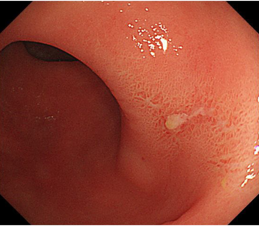
Figure 1 Colonoscopy examination showed redness and erosion on the rectal mucosa.
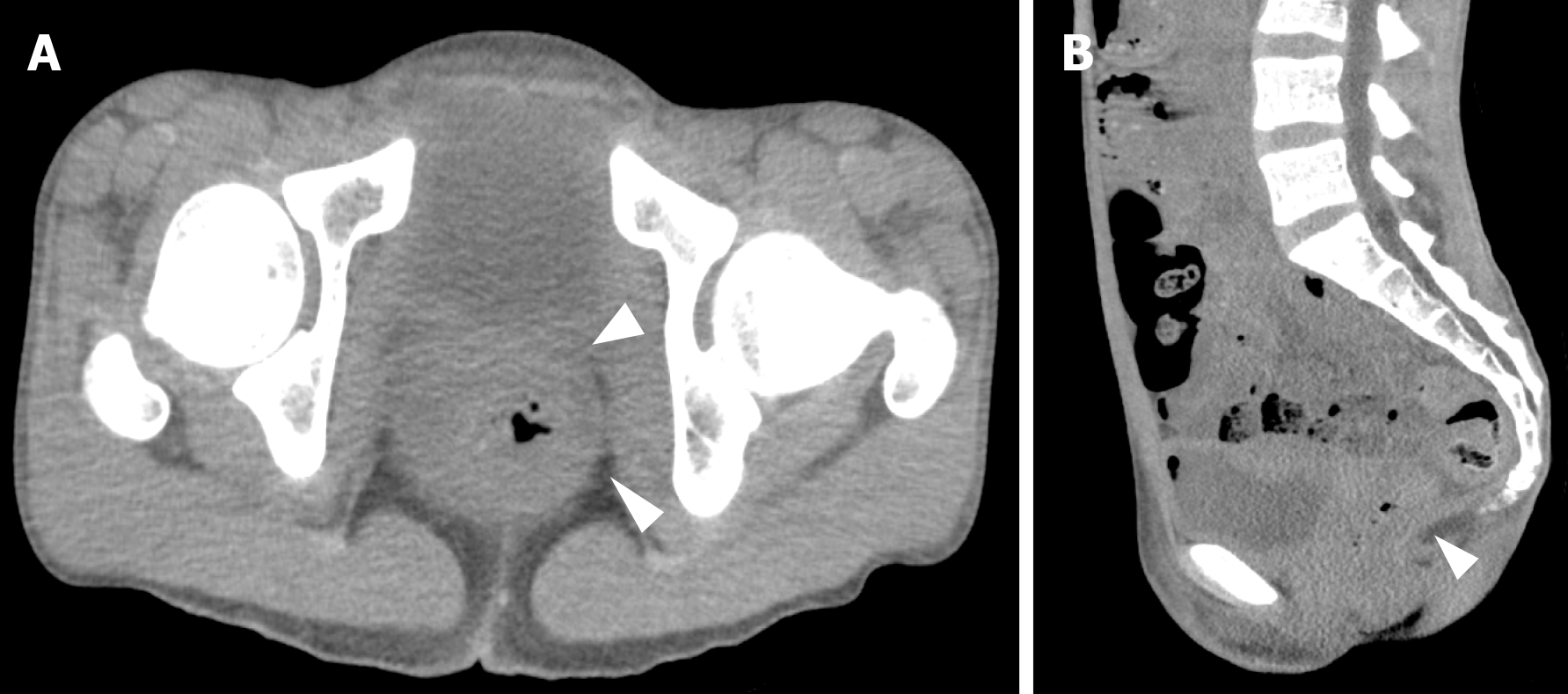
Figure 2 Computed tomography scans of the abdomen and pelvis.
A: Diffuse rectal wall thickening (arrowheads) was seen; B: The peritoneal reflection lay below the lower sacrum, accompanied by a thickened rectal wall.
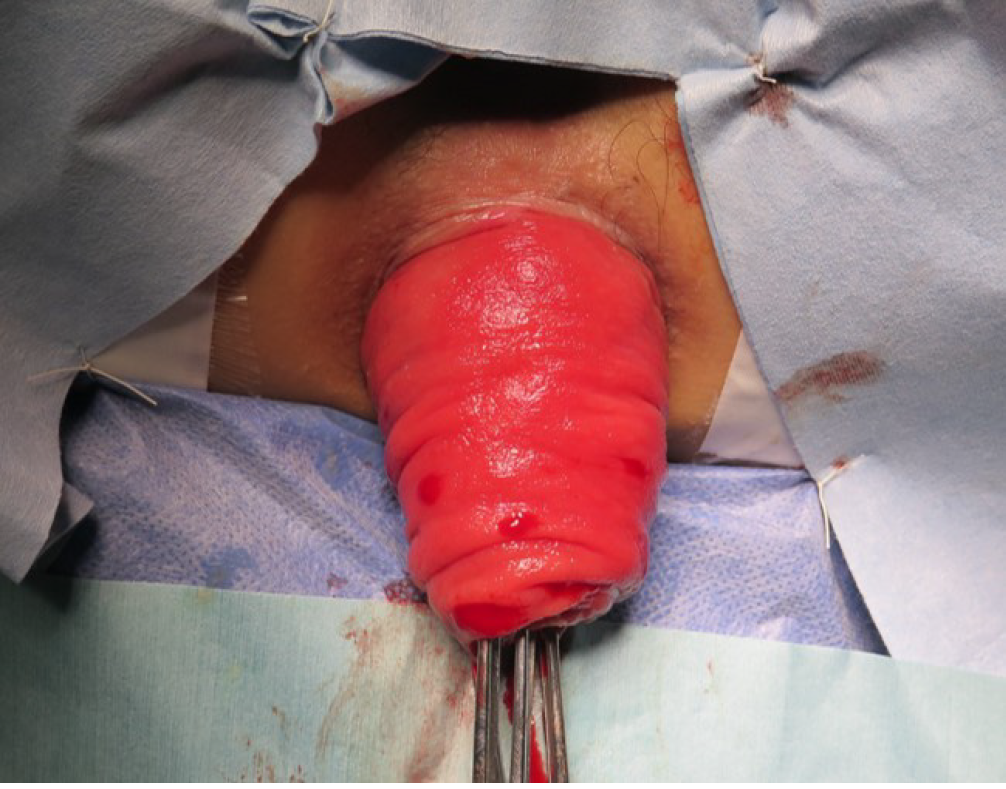
Figure 3 Preoperative appearance of the rectal prolapse.
A complete rectal prolapse, with a 10 cm-long reddish section of the rectum, is seen.
Figure 4 Intraoperative indocyanine green fluorescence imaging.
A: The proximal rectum (arrowhead) was pulled out through the anus [before indocyanine green (ICG) administration]; B: ICG fluorescence imaging of the proximal rectum (arrowhead), showing a visible blood supply to the rectum. The rectum was transected on the line with good blood flow (arrow); C: Completion of the Altemeier operation.
- Citation: Yamamoto T, Hyakudomi R, Takai K, Taniura T, Uchida Y, Ishitobi K, Hirahara N, Tajima Y. Altemeier perineal rectosigmoidectomy with indocyanine green fluorescence imaging for a female adolescent with complete rectal prolapse: A case report. World J Clin Cases 2021; 9(4): 847-853
- URL: https://www.wjgnet.com/2307-8960/full/v9/i4/847.htm
- DOI: https://dx.doi.org/10.12998/wjcc.v9.i4.847


