Copyright
©The Author(s) 2021.
World J Clin Cases. Feb 6, 2021; 9(4): 764-773
Published online Feb 6, 2021. doi: 10.12998/wjcc.v9.i4.764
Published online Feb 6, 2021. doi: 10.12998/wjcc.v9.i4.764
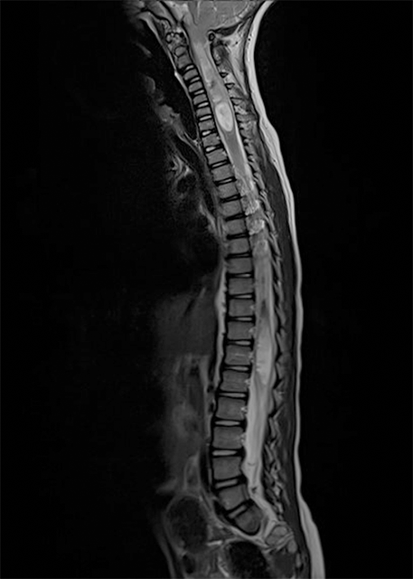
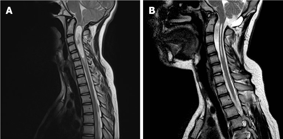
Figure 1 Chiari type 1 and associated holocord syringomyelia.
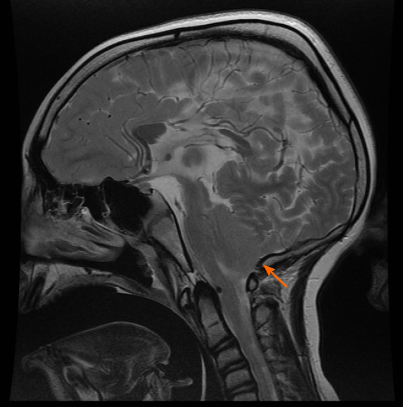
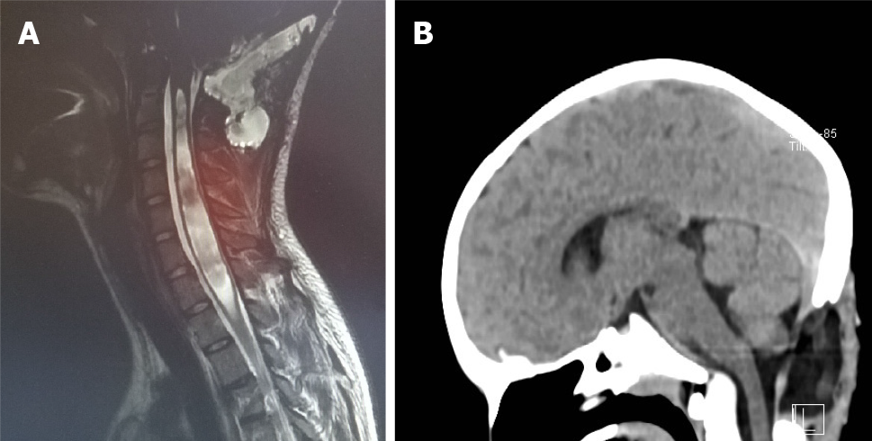
Figure 2 Chiari type 2 is characterized by a herniation of the tonsils, brainstem, vermis and also by corpus callosum agenesia, vermian agenesia, small posterior fossa, hydrocephalus and many other malformation of the central nervous system.
The arrow indicates a low-lying torcular, which impeded a wide occipital craniectomy at surgery.
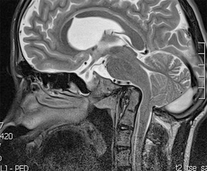
Figure 3 Basilar invagination associated to Chiari type 1.
In these cases, surgical treatment must take into account, beyond a craniocervical decompression, also a craniocervical fixation.
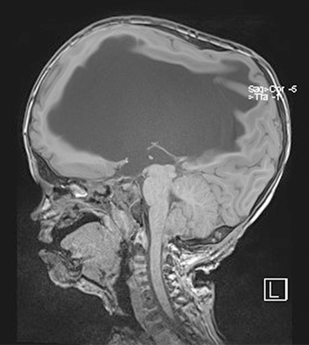
Figure 4 Chiari type 1 associated to a syndromic craniosynostosis (Pfeiffer syndrome).
There is also severe hydrocephalus, related to an intracranial venous hypertension.
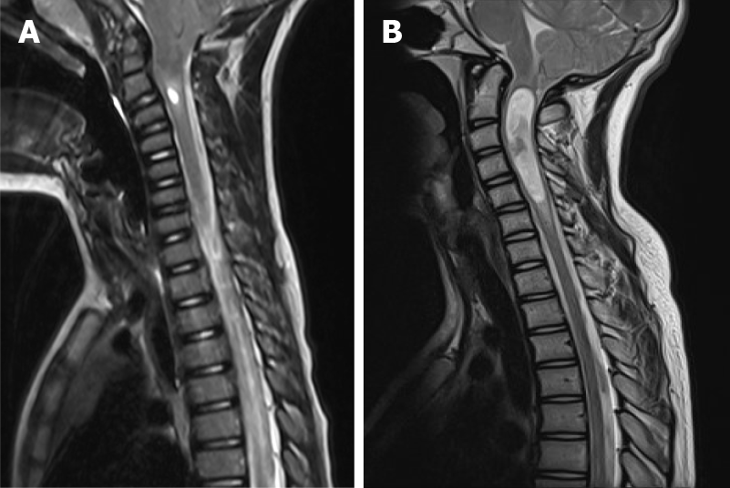
Figure 5 Two cases of syringomyelia caused by Chiari type 1 and limited to a limited segment of the spinal cord.
A and B: Cerebellar signs and symptoms are not present in Chiari type 1, since no eloquent neurological functions are located in the tonsils.
Figure 6 Hydromyelia, which indicated just a mild dilatation of the central spinal canal.
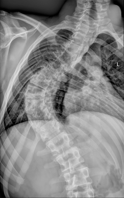
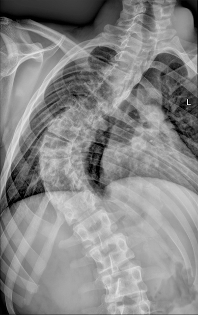
Figure 7 Mild or severe forms of scoliosis can be often found in Chiari malformations.
Figure 8 A case of successfully treated Chiari type 1 with craniocervical decompression.
A: A large syringomyelia before surgery; B: A large syringomyelia disappeared after the craniocervical decompression.
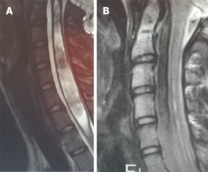
Figure 9 Another case of successful treatment of Chiari type 1 with craniocervical decompression.
A: The syringomyelia in the cervical spinal cord; B: A procedure that results in cervical spinal cord syringomyelia.
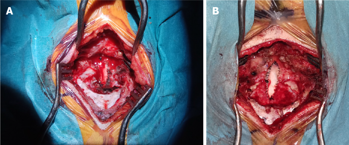
Figure 10 Two cases of craniocervical decompression.
A and B: There is a dural patch implanted in both cases. This allows an enlargement of the diameter of the dural sac at the craniocervical junction and consequently a larger space for cerebrospinal fluid circulation and rhomboencephalic structures.
Figure 11 Images show a pseudomeningocele, caused by a leak of cerebrospinal fluid in the epidural space.
A and B: The most common complications include injury to the vascular and neural structures, pseudomeningocele. This is a frequent complication of craniocervcal decompression.
- Citation: Spazzapan P, Bosnjak R, Prestor B, Velnar T. Chiari malformations in children: An overview. World J Clin Cases 2021; 9(4): 764-773
- URL: https://www.wjgnet.com/2307-8960/full/v9/i4/764.htm
- DOI: https://dx.doi.org/10.12998/wjcc.v9.i4.764


