Copyright
©The Author(s) 2021.
World J Clin Cases. Dec 26, 2021; 9(36): 11482-11486
Published online Dec 26, 2021. doi: 10.12998/wjcc.v9.i36.11482
Published online Dec 26, 2021. doi: 10.12998/wjcc.v9.i36.11482
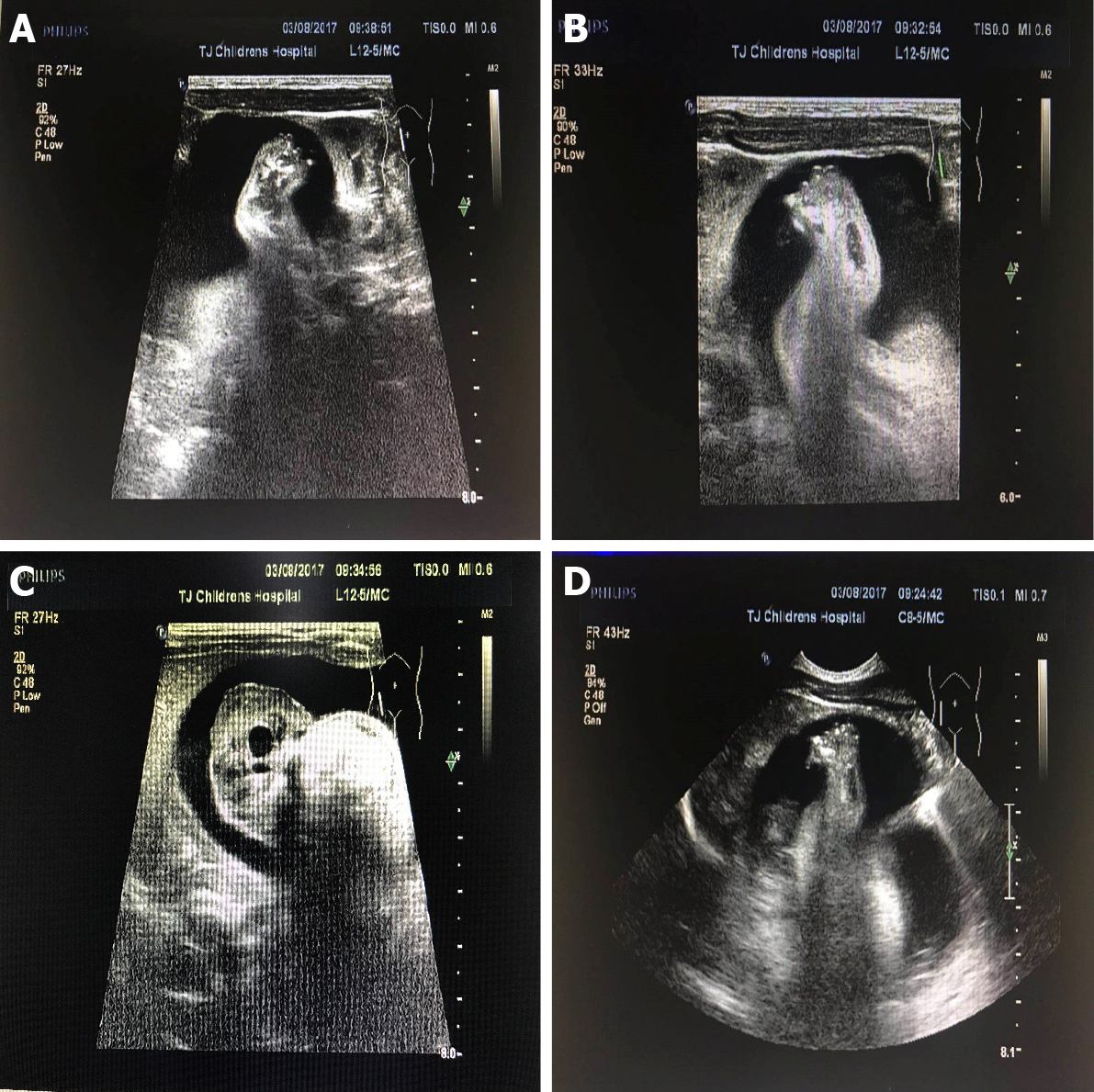
Figure 1 Feet and trunk of the fetus in fetu on ultrasound.
A, B: Feet; C, D: Trunk.
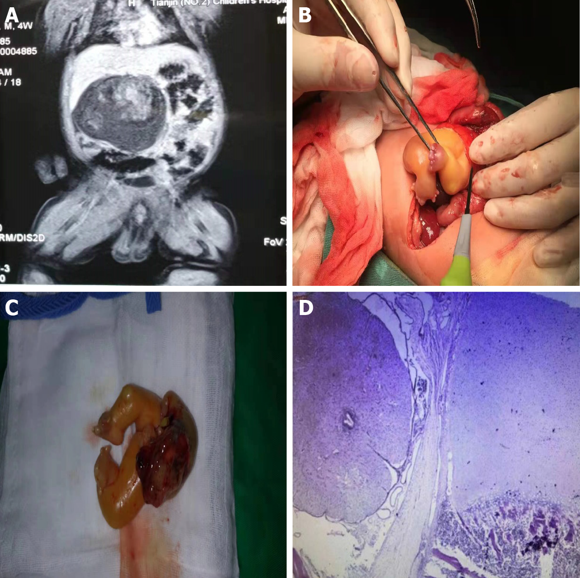
Figure 2 Images of the intestines and pathology of the mass.
A: The male infant’s intestines on magnetic resonance imaging; B, C: The feet and part of the abdominal cavity during the operation; D: Postoperative pathology of the mass.
- Citation: Xia B, Li DD, Wei HX, Zhang XX, Li RM, Chen J. Retroperitoneal parasitic fetus: A case report. World J Clin Cases 2021; 9(36): 11482-11486
- URL: https://www.wjgnet.com/2307-8960/full/v9/i36/11482.htm
- DOI: https://dx.doi.org/10.12998/wjcc.v9.i36.11482


